3K44 image
Deposition Date 2009-10-05
Release Date 2009-10-27
Last Version Date 2024-02-21
Entry Detail
PDB ID:
3K44
Title:
Crystal Structure of Drosophila melanogaster Pur-alpha
Biological Source:
Source Organism:
Host Organism:
Method Details:
Experimental Method:
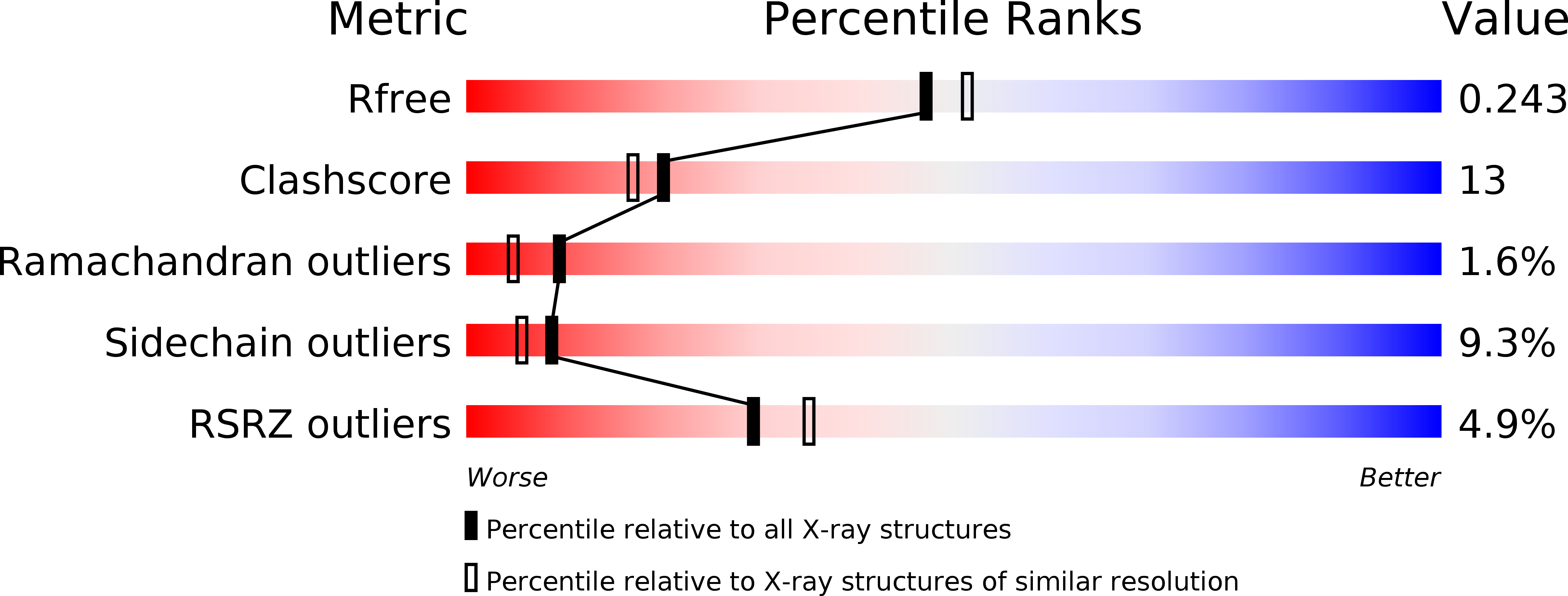
Resolution:
2.10 Å
R-Value Free:
0.24
R-Value Work:
0.22
R-Value Observed:
0.22
Space Group:
P 1 2 1
Macromolecular Entities
Polymer Type:polypeptide(L)
Molecule:Purine-rich binding protein-alpha, isoform B
Chain IDs:A, B, C, D
Chain Length:146
Number of Molecules:4
Biological Source:Drosophila melanogaster
Primary Citation
X-ray structure of Pur-alpha reveals a Whirly-like fold and an unusual nucleic-acid binding surface
Proc.Natl.Acad.Sci.USA 106 18521 18526 (2009)
PMID: 19846792 DOI: 10.1073/pnas.0907990106

Abstact

The PUR protein family is a distinct and highly conserved class that is characterized by its sequence-specific RNA- and DNA-binding. Its best-studied family member, Pur-alpha, acts as a transcriptional regulator, as host factor for viral replication, and as cofactor for mRNP localization in dendrites. Pur-alpha-deficient mice show severe neurologic defects and die after birth. Nucleic-acid binding by Pur-alpha is mediated by its central core region, for which no structural information is available. We determined the x-ray structure of residues 40 to 185 from Drosophila melanogaster Pur-alpha, which constitutes a major part of the core region. We found that this region contains two almost identical structural motifs, termed "PUR repeats," which interact with each other to form a PUR domain. DNA- and RNA-binding studies confirmed that PUR domains are indeed functional nucleic-acid binding domains. Database analysis show that PUR domains share a fold with the Whirly class of nucleic-acid binding proteins. Structural analysis combined with mutational studies suggest that a PUR domain binds nucleic acids through two independent surface regions involving concave beta-sheets. Structure-based sequence alignment revealed that the core region harbors a third PUR repeat at its C terminus. Subsequent characterization by small-angle x-ray scattering (SAXS) and size-exclusion chromatography indicated that PUR repeat III mediates dimerization of Pur-alpha. Surface envelopes calculated from SAXS data show that the Pur-alpha dimer consisting of repeats I to III is arranged in a Z-like shape. This unexpected domain organization of the entire core domain of Pur-alpha has direct implications for ssDNA/ssRNA and dsDNA binding.

Legend

Protein

Chemical

Disease

Primary Citation of related structures