
Deposition Date
2009-10-04
Release Date
2010-07-14
Last Version Date
2023-11-01
Entry Detail
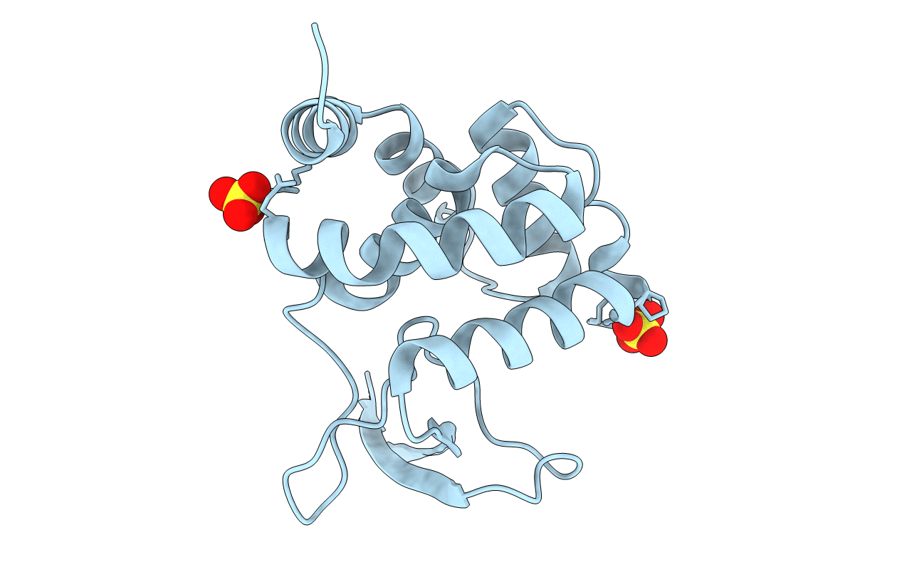
PDB ID:
3K3T
Keywords:
Title:
E185A mutant of peptidoglycan hydrolase from Sphingomonas sp. A1
Biological Source:
Source Organism(s):
Sphingomonas sp. A1 (Taxon ID: 90322)
Expression System(s):
Method Details:
Experimental Method:
Resolution:
1.75 Å
R-Value Free:
0.22
R-Value Work:
0.19
R-Value Observed:
0.19
Space Group:
P 43 21 2
