Deposition Date
2009-10-02
Release Date
2009-11-17
Last Version Date
2024-02-21
Entry Detail
PDB ID:
3K3F
Keywords:
Title:
Crystal Structure of the Urea Transporter from Desulfovibrio Vulgaris
Biological Source:
Source Organism(s):
Desulfovibrio vulgaris (Taxon ID: 882)
Expression System(s):
Method Details:
Experimental Method:
Resolution:
2.30 Å
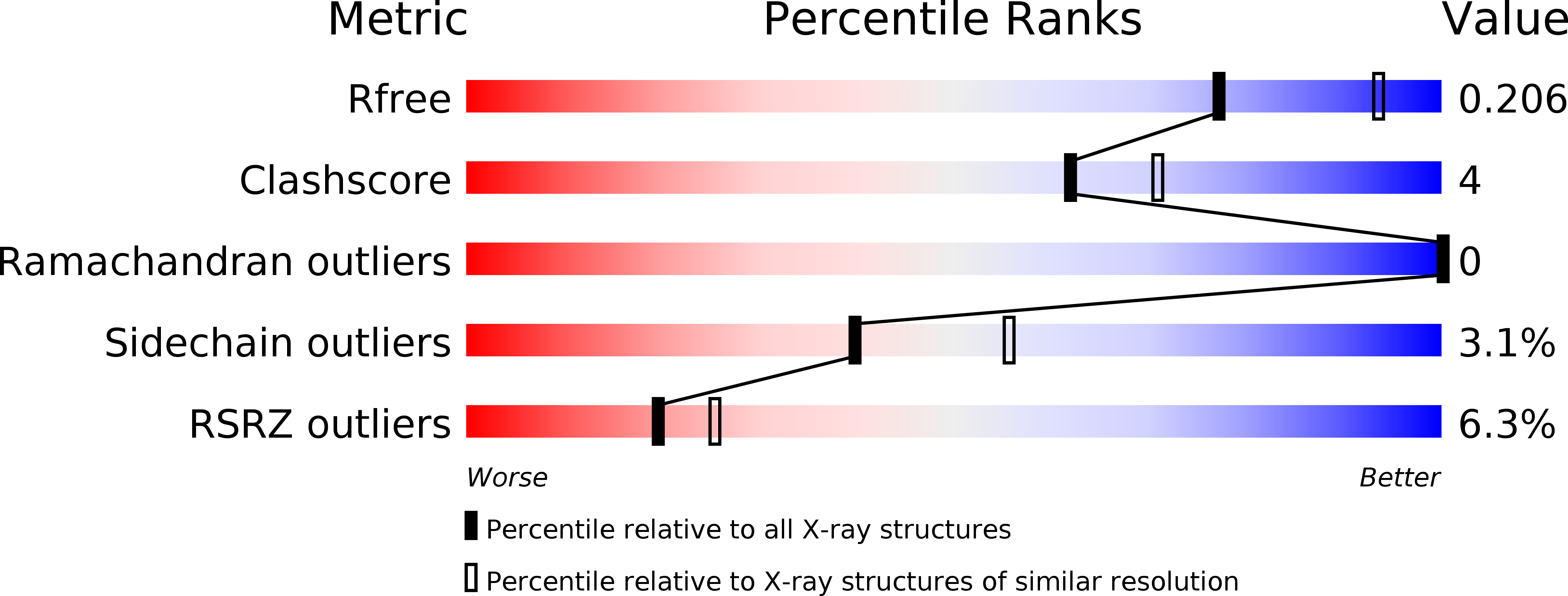
R-Value Free:
0.20
R-Value Work:
0.17
R-Value Observed:
0.18
Space Group:
P 63