3K2O image
Deposition Date 2009-09-30
Release Date 2009-11-03
Last Version Date 2024-11-13
Entry Detail
PDB ID:
3K2O
Keywords:
Title:
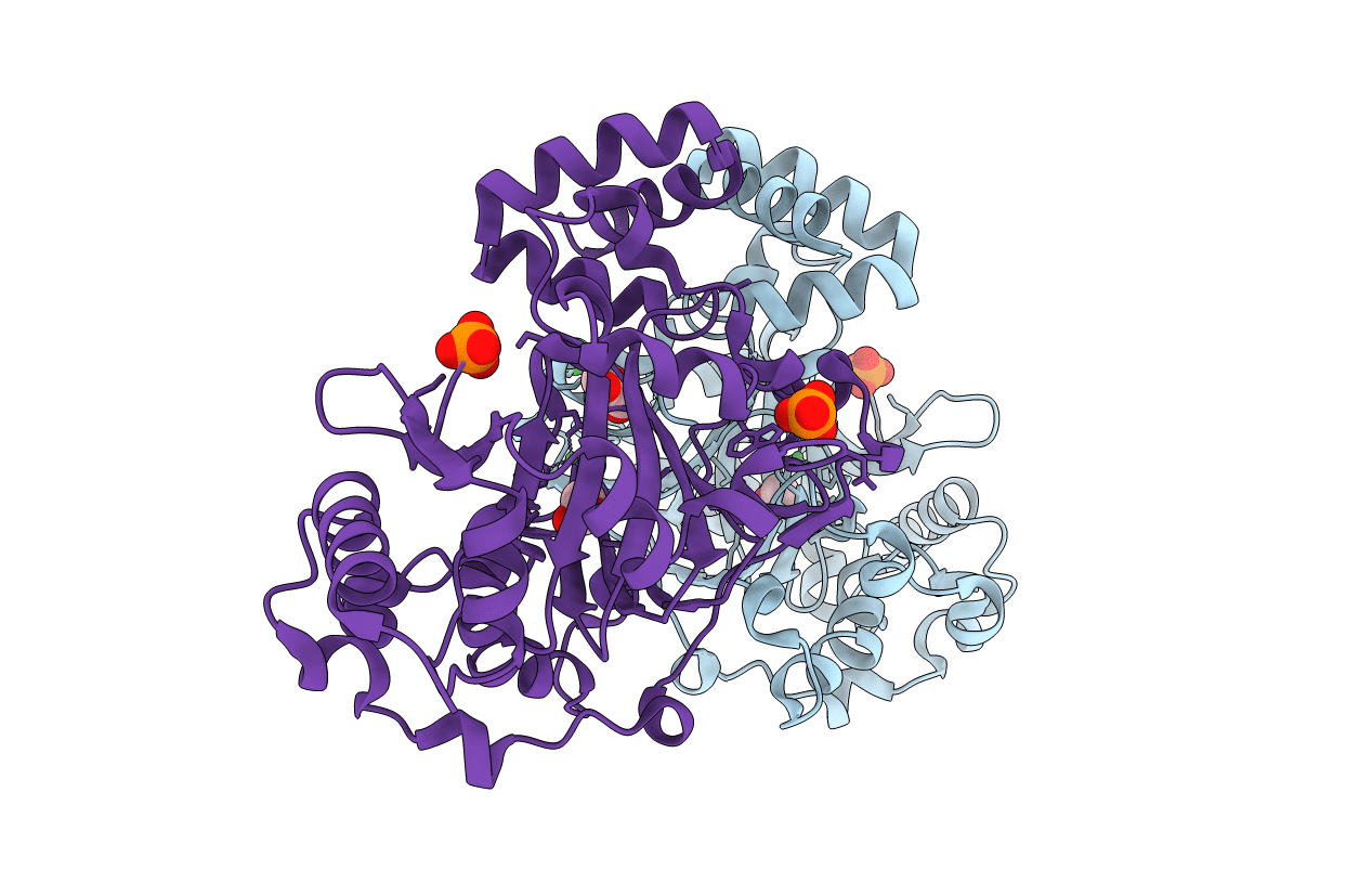
Structure of an oxygenase
Biological Source:
Source Organism:
Homo sapiens (Taxon ID: 9606)
Host Organism:
Method Details:
Experimental Method:
Resolution:
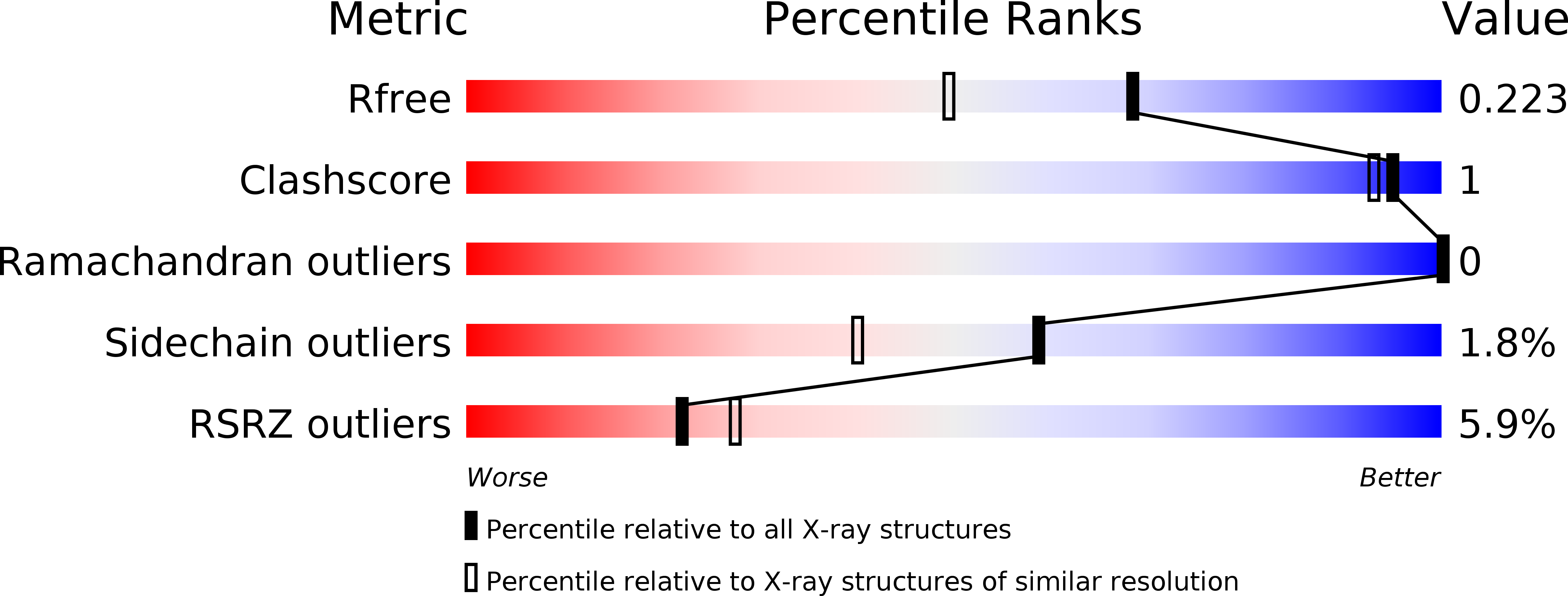
1.75 Å
R-Value Free:
0.22
R-Value Work:
0.19
R-Value Observed:
0.19
Space Group:
P 1 21 1
Macromolecular Entities
Structures with similar UniProt ID
Protein Blast
Polymer Type:polypeptide(L)
Molecule:Bifunctional arginine demethylase and lysyl-hydroxylase JMJD6
Gene (Uniprot):JMJD6
Chain IDs:A, B
Chain Length:336
Number of Molecules:2
Biological Source:Homo sapiens
Modified Residue
Compound ID Chain ID Parent Comp ID Details 2D Image
MSE A MET SELENOMETHIONINE
Primary Citation
Crystal Structure of the 2-Oxoglutarate- and Fe(II)-Dependent Lysyl Hydroxylase JMJD6.
J.Mol.Biol. 401 211 222 (2010)
PMID: 20685276 DOI: 10.1016/j.jmb.2010.05.054

Abstact

Lysyl and prolyl hydroxylations are well-known post-translational modifications to animal and plant proteins with extracellular roles. More recent work has indicated that the hydroxylation of intracellular animal proteins may be common. JMJD6 catalyses the iron- and 2-oxoglutarate-dependent hydroxylation of lysyl residues in arginine-serine-rich domains of RNA-splicing-related proteins. We report crystallographic studies on the catalytic domain of JMJD6 in complex with Ni(II) substituting for Fe(II). Together with mutational studies, the structural data suggest how JMJD6 binds its lysyl residues such that it can catalyse C-5 hydroxylation rather than N(varepsilon)-demethylation, as for analogous enzymes.

Legend

Protein

Chemical

Disease

Primary Citation of related structures