3K1A image
Deposition Date 2009-09-26
Release Date 2010-02-16
Last Version Date 2023-09-06
Entry Detail
PDB ID:
3K1A
Keywords:
Title:
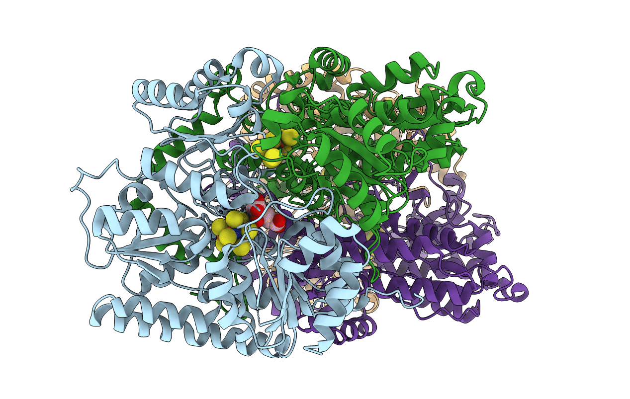
Insights into substrate binding at FeMo-cofactor in nitrogenase from the structure of an alpha-70Ile MoFe protein variant
Biological Source:
Source Organism:
Host Organism:
Method Details:
Experimental Method:
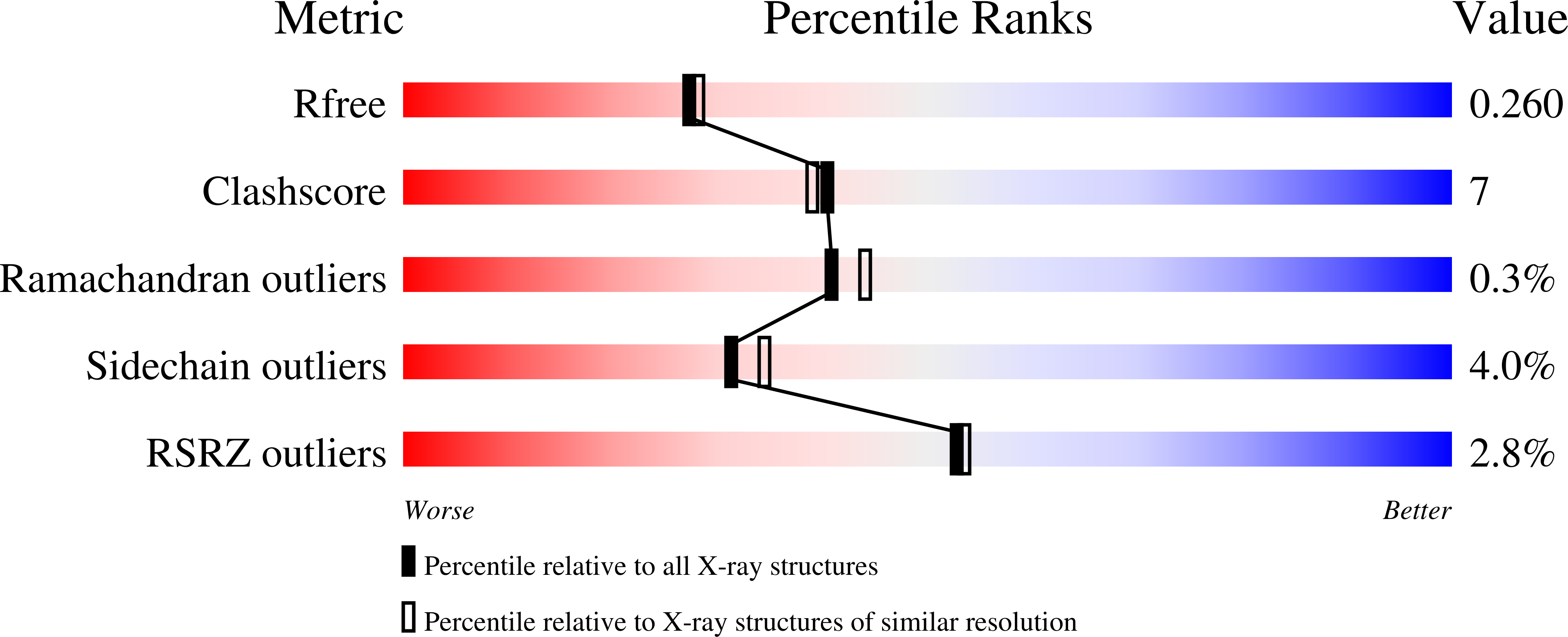
Resolution:
2.23 Å
R-Value Free:
0.25
R-Value Work:
0.20
R-Value Observed:
0.20
Space Group:
P 1 21 1
Macromolecular Entities
Polymer Type:polypeptide(L)
Molecule:Nitrogenase molybdenum-iron protein alpha chain
Gene (Uniprot):nifD
Mutations:V70I
Chain IDs:A, C
Chain Length:491
Number of Molecules:2
Biological Source:Azotobacter vinelandii
Polymer Type:polypeptide(L)
Molecule:Nitrogenase molybdenum-iron protein beta chain
Gene (Uniprot):nifK
Chain IDs:B, D
Chain Length:522
Number of Molecules:2
Biological Source:Azotobacter vinelandii
Primary Citation
Insights into substrate binding at FeMo-cofactor in nitrogenase from the structure of an alpha-70(Ile) MoFe protein variant
J.Inorg.Biochem. 104 385 389 (2010)
PMID: 20022118 DOI: 10.1016/j.jinorgbio.2009.11.009

Abstact

The X-ray crystal structure is presented for a nitrogenase MoFe protein where the alpha subunit residue at position 70 (alpha-70(Val)) has been substituted by the amino acid isoleucine (alpha-70(Ile)). Substitution of alpha-70(Val) by alpha-70(Ile) results in a MoFe protein that is hampered in its ability to reduce a range of substrates including acetylene and N(2), yet retains normal proton reduction activity. The 2.3A structure of the alpha-70(Ile) MoFe protein is compared to the alpha-70(Val) wild-type MoFe protein, revealing that the delta methyl group of alpha-70(Val) is positioned over Fe6 within the active site FeMo-cofactor. This work provides strong crystallographic support for the previously proposed model that substrates bind and are reduced at a single 4Fe-4S face of the FeMo-cofactor and that when alpha-70(Val) is substituted by alpha-70(Ile) access of substrates to Fe6 of this face is effectively blocked. Furthermore the detailed examination of the structure provides the basis for understanding the ability to trap and characterize hydrides in the variant, contributing significantly to our understanding of substrate access and substrate reduction at the FeMo-cofactor active site of nitrogenase.

Legend

Protein

Chemical

Disease

Primary Citation of related structures