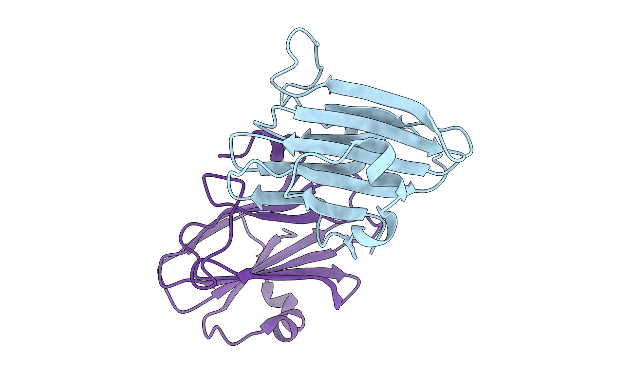
3JYB image
Entry Detail
PDB ID:
3JYB
Keywords:
Title:
Crystal Structure of the RetS periplasmic domain
Biological Source:
Source Organism:
Host Organism:
PDB Version:
Deposition Date:
2009-09-21
Release Date:
2010-02-16
Method Details:
Experimental Method:
Resolution:
2.04 Å
R-Value Free:
0.23
R-Value Work:
0.19
R-Value Observed:
0.20
Space Group:
P 21 21 21
Macromolecular Entities
Polymer Type:polypeptide(L)
Description:Sensor protein
Chain IDs:A, B
Chain Length:145
Number of Molecules:2
Biological Source:Pseudomonas aeruginosa
Modified Residue
Compound ID Chain ID Parent Comp ID Details 2D Image
MSE A MET SELENOMETHIONINE
Primary Citation
Crystal structure and oligomeric state of the RetS signaling kinase sensory domain.
Proteins 78 1631 1640 (2010)
PMID: 20112417 DOI: 10.1002/prot.22679

Abstact

The opportunistic pathogen Pseudomonas aeruginosa may cause both acute and chronic-persistent infections in predisposed individuals. Acute infections require the presence of a functional type III secretion system (T3SS), whereas chronic P. aeruginosa infections are characterized by the formation of drug-resistant biofilms. The T3SS and biofilm formation are reciprocally regulated by the signaling kinases LadS, RetS, and GacS. RetS downregulates biofilm formation and upregulates expression of the T3SS through a unique mechanism. RetS forms a heterodimeric complex with GacS and thus prevents GacS autophosphorylation and downstream signaling. The signals that regulate RetS are not known but RetS possesses a distinctive periplasmic sensor domain that is believed to serve as receptor for the regulatory ligand. We have determined the crystal structure of the RetS sensory domain at 2.0 A resolution. The structure closely resembles those of carbohydrate binding modules of other proteins, suggesting that the elusive ligands are likely carbohydrate moieties. In addition to the conserved beta-sandwich structure, the sensory domain features two alpha helices which create a unique surface topology. Protein-protein crosslinking and fluorescence energy transfer experiments also revealed that the sensory domain dimerizes with a dissociation constant of K(d) = 580 +/- 50 nM, a result with interesting implications for our understanding of the underlying signaling mechanism.

Legend

Protein

Chemical

Disease

Primary Citation of related structures