3JWB image
Deposition Date 2009-09-18
Release Date 2010-09-08
Last Version Date 2023-11-22
Entry Detail
PDB ID:
3JWB
Keywords:
Title:
Crystal structure of L-methionine gamma-lyase from Citrobacter freundii with norleucine
Biological Source:
Source Organism:
Host Organism:
Method Details:
Experimental Method:
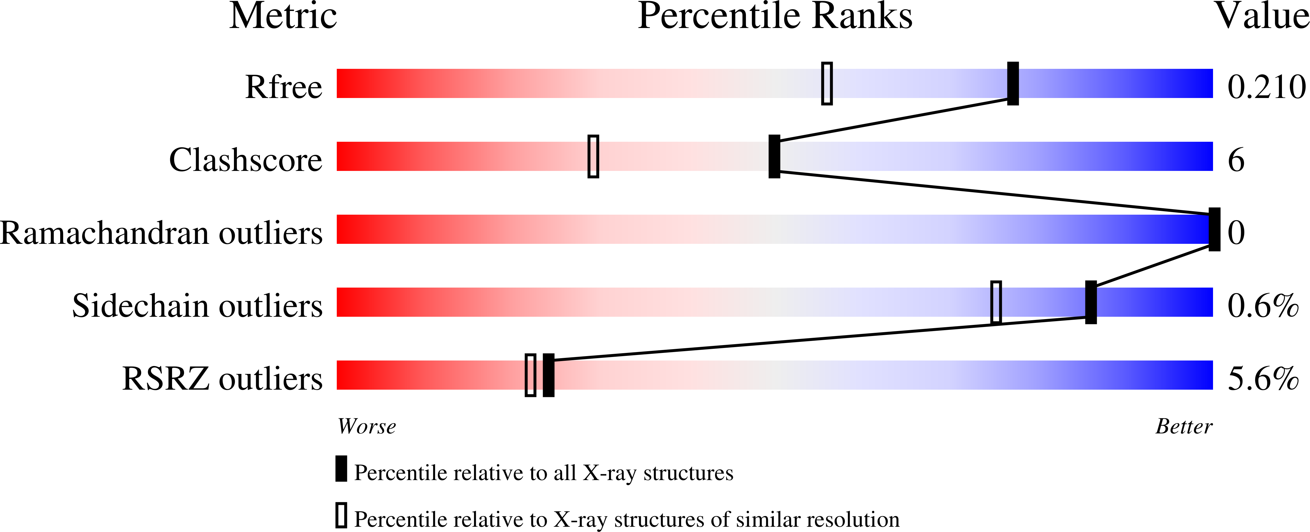
Resolution:
1.63 Å
R-Value Free:
0.21
R-Value Work:
0.15
R-Value Observed:
0.16
Space Group:
I 2 2 2
Macromolecular Entities
Polymer Type:polypeptide(L)
Molecule:Methionine gamma-lyase
Gene (Uniprot):megL
Chain IDs:A
Chain Length:398
Number of Molecules:1
Biological Source:Citrobacter freundii
Modified Residue
Compound ID Chain ID Parent Comp ID Details 2D Image
CSO A CYS S-HYDROXYCYSTEINE
LLP A LYS ?
Primary Citation
Three-dimensional structures of noncovalent complexes of Citrobacter freundii methionine gamma-lyase with substrates.
Biochemistry Mosc. 76 564 570 (2011)
PMID: 21639836 DOI: 10.1134/S0006297911050063

Abstact

Crystal structures of Citrobacter freundii methionine γ-lyase complexes with the substrates of γ- (L-1-amino-3-methylthiopropylphosphinic acid) and β- (S-ethyl-L-cysteine) elimination reactions and the competitive inhibitor L-norleucine have been determined at 1.45, 1.8, and 1.63 Å resolution, respectively. All three amino acids occupy the active site of the enzyme but do not form a covalent bond with pyridoxal 5'-phosphate. Hydrophobic interactions between the active site residues and the side groups of the substrates and the inhibitor are supposed to cause noncovalent binding. Arg374 and Ser339 are involved in the binding of carboxyl groups of the substrates and the inhibitor. The hydroxyl of Tyr113 is a potential acceptor of a proton from the amino groups of the amino acids.

Legend

Protein

Chemical

Disease

Primary Citation of related structures