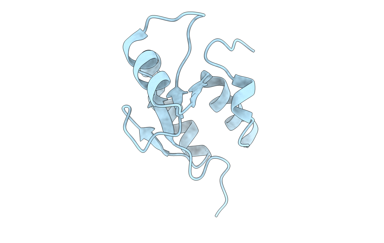
3JVE image
Entry Detail
PDB ID:
3JVE
Keywords:
Title:
Crystal Structure of the Sixth BRCT Domain of TopBP1
Biological Source:
Source Organism:
Host Organism:
PDB Version:
Deposition Date:
2009-09-16
Release Date:
2010-01-19
Method Details:
Experimental Method:
Resolution:
1.34 Å
R-Value Free:
0.17
R-Value Work:
0.15
R-Value Observed:
0.15
Space Group:
P 21 21 21
Macromolecular Entities
Polymer Type:polypeptide(L)
Description:DNA topoisomerase 2-binding protein 1
Chain IDs:A
Chain Length:109
Number of Molecules:1
Biological Source:Homo sapiens
Primary Citation
Insights from the crystal structure of the sixth BRCT domain of topoisomerase IIbeta binding protein 1.
Protein Sci. 19 162 167 (2010)
PMID: 19937654 DOI: 10.1002/pro.290

Abstact

Topoisomerase IIbeta binding protein 1 (TopBP1) is a major player in the DNA damage response and interacts with a number of protein partners via its eight BRCA1 carboxy-terminal (BRCT) domains. In particular, the sixth BRCT domain of TopBP1 has been implicated in binding to the phosphorylated transcription factor, E2F1, and poly(ADP-ribose) polymerase 1 (PARP-1), where the latter interaction is responsible for the poly(ADP-ribosyl)ation of TopBP1. To gain a better understanding of the nature of TopBP1 BRCT6 interactions, we solved the crystal structure of BRCT6 to 1.34 A. The crystal structure reveals a degenerate phospho-peptide binding pocket and lacks conserved hydrophobic residues involved in packing of tandem BRCT repeats, which, together with results from phospho-peptide binding studies, strongly suggest that TopBP1 BRCT6 independently does not function as a phospho-peptide binding domain. We further provide insight into poly(ADP-ribose) binding and sites of potential modification by PARP-1.

Legend

Protein

Chemical

Disease

Primary Citation of related structures