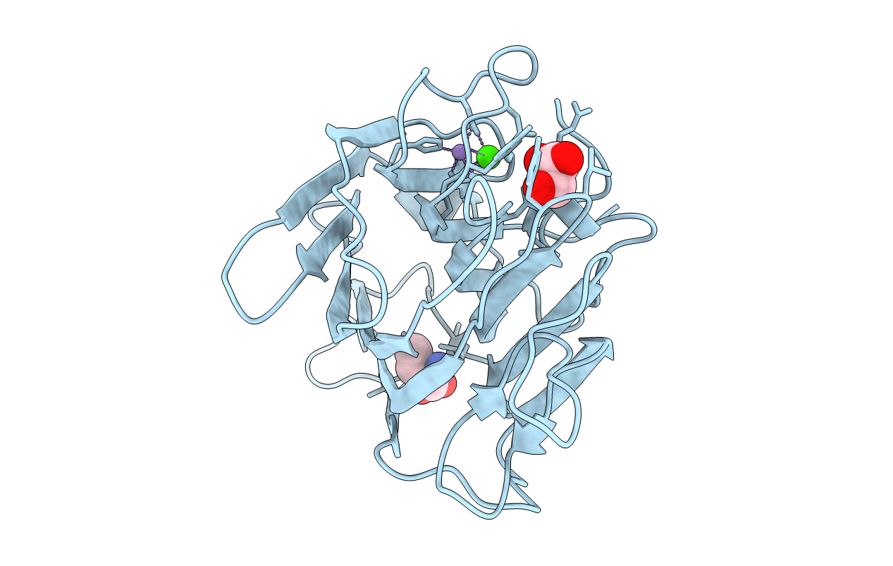
3JU9 image
Entry Detail
PDB ID:
3JU9
Title:
Crystal structure of a lectin from Canavalia brasiliensis seed (ConBr) complexed with alpha-aminobutyric acid
Biological Source:
Source Organism:
PDB Version:
Deposition Date:
2009-09-14
Release Date:
2010-12-08
Method Details:
Experimental Method:
Resolution:
2.10 Å
R-Value Free:
0.25
R-Value Work:
0.20
R-Value Observed:
0.20
Space Group:
I 2 2 2
Macromolecular Entities
Polymer Type:polypeptide(L)
Description:Concanavalin-Br
Chain IDs:A
Chain Length:237
Number of Molecules:1
Biological Source:Canavalia brasiliensis
Primary Citation
Structural analysis of ConBr reveals molecular correlation between the carbohydrate recognition domain and endothelial NO synthase activation.
Biochem.Biophys.Res.Commun. 408 566 570 (2011)
PMID: 21530490 DOI: 10.1016/j.bbrc.2011.04.061

Abstact

Diocleinae lectins are highly homologous in their primary structure which features metal binding sites and a carbohydrate recognition domain (CRD). Differences in the biological activity of legume lectins have been widely investigated using hemagglutination inhibition assays, isothermal titration microcalorimetry and co-crystallization with mono- and oligosaccharides. Here we report a new lectin crystal structure (ConBr) extracted from seeds of Canavalia brasiliensis, predict dimannoside binding by docking, identify the α-aminobutyric acid (Abu) binding pocket and compare the CRD of ConBr to that of homologous lectins. Based on the hypothesis that the carbohydrate affinity of lectins depends on CRD configuration, the relationship between tridimensional structure and endothelial NO synthase activation was used to clarify differences in biological activity. Our study established a correlation between the position of CRD amino acid side chains and the stimulation of NO release from endothelium.

Legend

Protein

Chemical

Disease

Primary Citation of related structures