3JTR image
Deposition Date 2009-09-14
Release Date 2010-01-26
Last Version Date 2023-11-01
Entry Detail
PDB ID:
3JTR
Keywords:
Title:
Mutations in Cephalosporin Acylase Affecting Stability and Autoproteolysis
Biological Source:
Source Organism:
Host Organism:
Method Details:
Experimental Method:
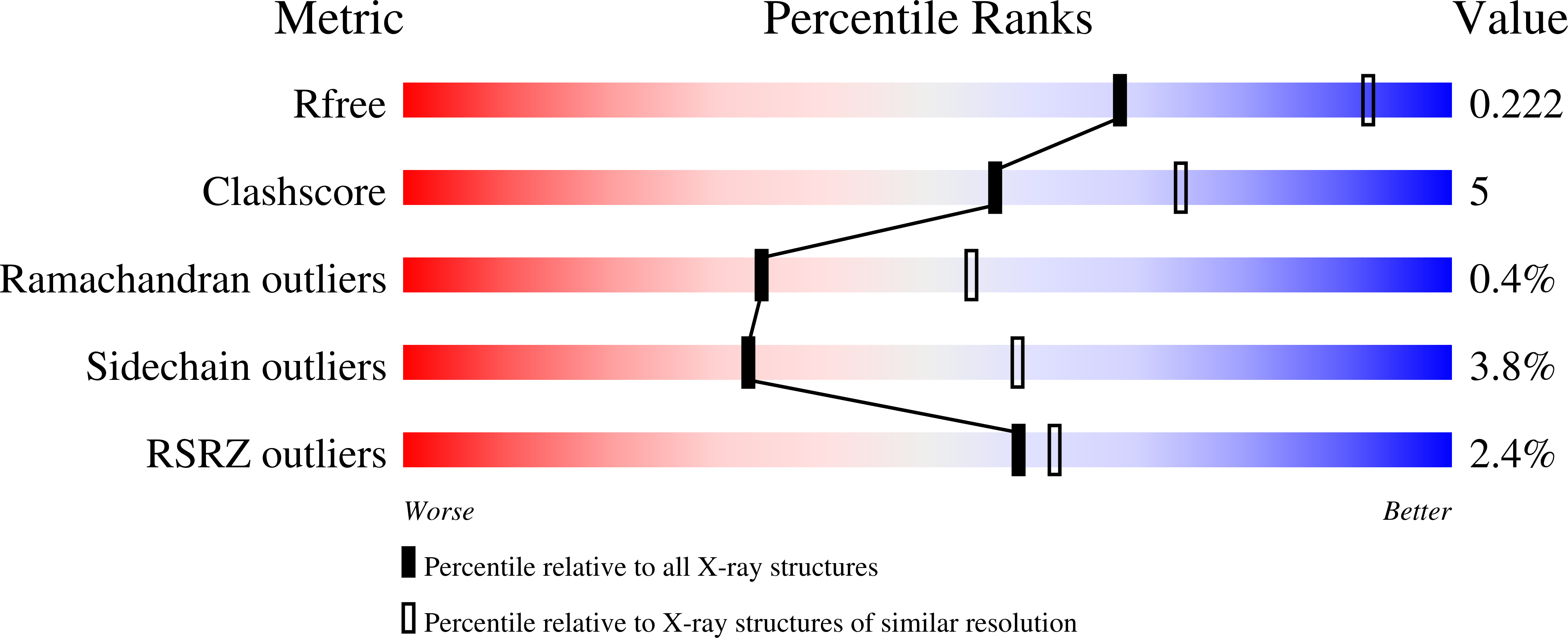
Resolution:
2.50 Å
R-Value Free:
0.22
R-Value Work:
0.18
R-Value Observed:
0.18
Space Group:
P 41 21 2
Macromolecular Entities
Structures with similar UniProt ID
Protein Blast
Polymer Type:polypeptide(L)
Molecule:Glutaryl 7-aminocephalosporanic acid acylase
Mutagens:Q168P
Chain IDs:A
Chain Length:169
Number of Molecules:1
Biological Source:Pseudomonas sp.
Structures with similar UniProt ID
Protein Blast
Polymer Type:polypeptide(L)
Molecule:Glutaryl 7-aminocephalosporanic acid acylase
Chain IDs:B
Chain Length:528
Number of Molecules:1
Biological Source:Pseudomonas sp.
Ligand Molecules
Primary Citation
Structural features of cephalosporin acylase reveal the basis of autocatalytic activation.
Biochem.Biophys.Res.Commun. 390 342 348 (2009)
PMID: 19800869 DOI: 10.1016/j.bbrc.2009.09.134

Abstact

Cephalosporin acylase (CA), a member of the N-terminal nucleophile hydrolase family, is activated through two steps of intramolecular autoproteolysis, the first mediated by a serine residue, and the second by a glutamate, which releases the pro-segment and produces an active enzyme. In this study, we have determined the crystal structures of mutants which could affect primary or secondary auto-cleavage and of sequential intermediates of a slow-processing mutant at 2.0-2.5A resolutions. The pro-segments of the mutants undergo dynamic conformational changes during activation and adopt surprisingly different loop conformations from one another. However, the autoproteolytic site was found to form a catalytically competent conformation with a solvent water molecule, which was essentially conserved in the CA mutants.

Legend

Protein

Chemical

Disease

Primary Citation of related structures