
Deposition Date
2009-09-08
Release Date
2010-08-18
Last Version Date
2024-04-03
Entry Detail
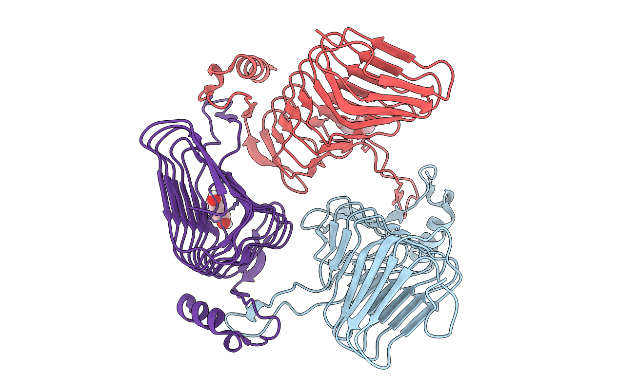
PDB ID:
3JQY
Keywords:
Title:
Crystal Structure of the polySia specific acetyltransferase NeuO
Biological Source:
Source Organism(s):
Escherichia coli (Taxon ID: 562)
Expression System(s):
Method Details:
Experimental Method:
Resolution:
1.70 Å
R-Value Free:
0.19
R-Value Work:
0.16
R-Value Observed:
0.16
Space Group:
P 1 21 1
