3JQO image
Deposition Date 2009-09-07
Release Date 2009-12-01
Last Version Date 2024-10-16
Entry Detail
PDB ID:
3JQO
Title:
Crystal structure of the outer membrane complex of a type IV secretion system
Biological Source:
Source Organism:
Host Organism:
Method Details:
Experimental Method:
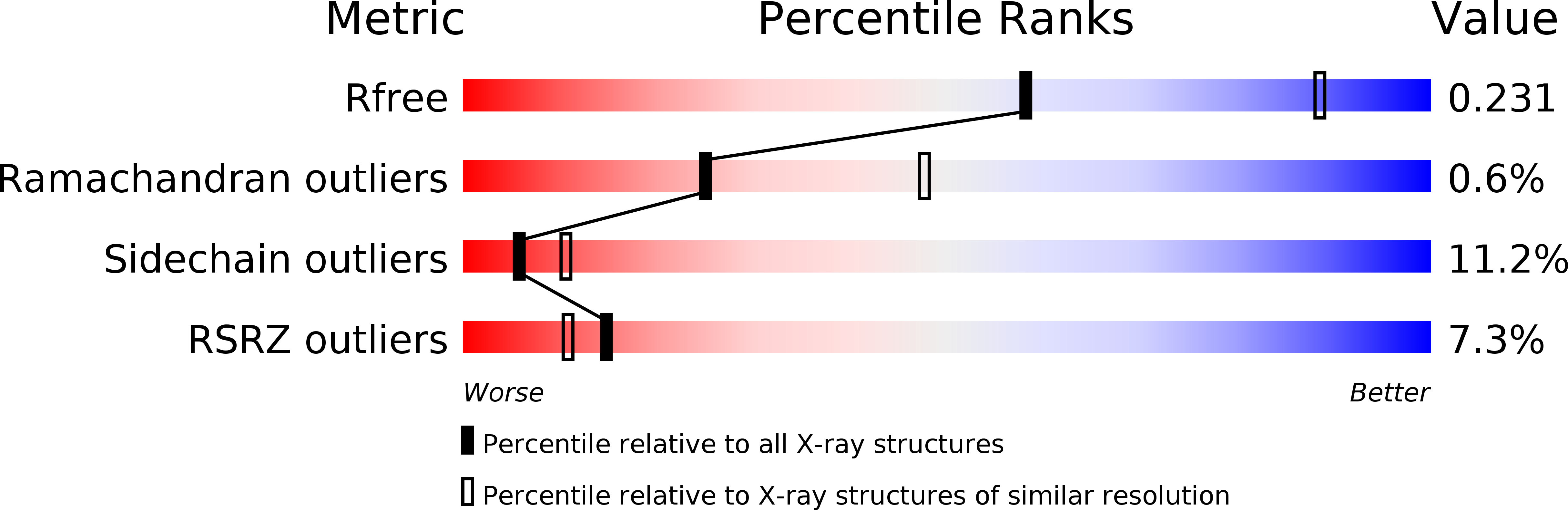
Resolution:
2.60 Å
R-Value Free:
0.25
R-Value Work:
0.22
R-Value Observed:
0.22
Space Group:
P 21 21 2
Macromolecular Entities
Polymer Type:polypeptide(L)
Molecule:TraF protein
Gene (Uniprot):traF
Chain IDs:A, D, G, J, M, P, S, V, Y, BA (auth: b), EA (auth: e), HA (auth: h), KA (auth: k), NA (auth: n)
Chain Length:227
Number of Molecules:14
Biological Source:Escherichia coli
Polymer Type:polypeptide(L)
Molecule:TraO protein
Gene (Uniprot):traO
Chain IDs:B, E, H, K, N, Q, T, W, Z, CA (auth: c), FA (auth: f), IA (auth: i), LA (auth: l), OA (auth: o)
Chain Length:227
Number of Molecules:14
Biological Source:Escherichia coli
Polymer Type:polypeptide(L)
Molecule:TraN protein
Gene (Uniprot):traN
Chain IDs:C, F, I, L, O, R, U, X, AA (auth: a), DA (auth: d), GA (auth: g), JA (auth: j), MA (auth: m), PA (auth: p)
Chain Length:227
Number of Molecules:14
Biological Source:Escherichia coli
Modified Residue
Compound ID Chain ID Parent Comp ID Details 2D Image
MSE A MET SELENOMETHIONINE
Primary Citation
Structure of the outer membrane complex of a type IV secretion system
Nature 462 1011 1015 (2009)
PMID: 19946264 DOI: 10.1038/nature08588

Abstact

Type IV secretion systems are secretion nanomachines spanning the two membranes of Gram-negative bacteria. Three proteins, VirB7, VirB9 and VirB10, assemble into a 1.05 megadalton (MDa) core spanning the inner and outer membranes. This core consists of 14 copies of each of the proteins and forms two layers, the I and O layers, inserting in the inner and outer membrane, respectively. Here we present the crystal structure of a approximately 0.6 MDa outer-membrane complex containing the entire O layer. This structure is the largest determined for an outer-membrane channel and is unprecedented in being composed of three proteins. Unexpectedly, this structure identifies VirB10 as the outer-membrane channel with a unique hydrophobic double-helical transmembrane region. This structure establishes VirB10 as the only known protein crossing both membranes of Gram-negative bacteria. Comparison of the cryo-electron microscopy (cryo-EM) and crystallographic structures points to conformational changes regulating channel opening and closing.

Legend

Protein

Chemical

Disease

Primary Citation of related structures