3JPW image
Deposition Date 2009-09-04
Release Date 2009-12-08
Last Version Date 2024-10-30
Entry Detail
PDB ID:
3JPW
Title:
Crystal structure of amino terminal domain of the NMDA receptor subunit NR2B
Biological Source:
Source Organism:
Host Organism:
Method Details:
Experimental Method:
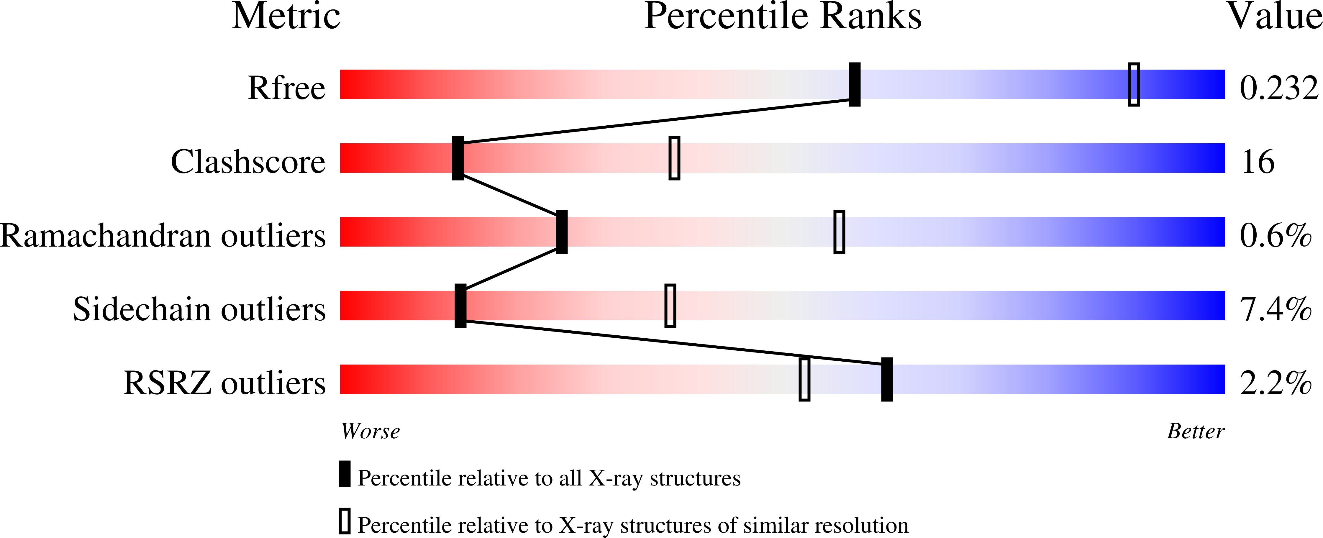
Resolution:
2.80 Å
R-Value Free:
0.23
R-Value Work:
0.19
R-Value Observed:
0.19
Space Group:
P 31 2 1
Macromolecular Entities
Polymer Type:polypeptide(L)
Molecule:Glutamate [NMDA] receptor subunit epsilon-2
Gene (Uniprot):Grin2b
Mutations:N348D
Chain IDs:A
Chain Length:363
Number of Molecules:1
Biological Source:Rattus norvegicus
Modified Residue
Compound ID Chain ID Parent Comp ID Details 2D Image
ASN A ASN GLYCOSYLATION SITE
Primary Citation
Structure of the zinc-bound amino-terminal domain of the NMDA receptor NR2B subunit.
Embo J. 28 3910 3920 (2009)
PMID: 19910922 DOI: 10.1038/emboj.2009.338

Abstact

N-methyl-D-aspartate (NMDA) receptors belong to the family of ionotropic glutamate receptors (iGluRs) that mediate the majority of fast excitatory synaptic transmission in the mammalian brain. One of the hallmarks for the function of NMDA receptors is that their ion channel activity is allosterically regulated by binding of modulator compounds to the extracellular amino-terminal domain (ATD) distinct from the L-glutamate-binding domain. The molecular basis for the ATD-mediated allosteric regulation has been enigmatic because of a complete lack of structural information on NMDA receptor ATDs. Here, we report the crystal structures of ATD from the NR2B NMDA receptor subunit in the zinc-free and zinc-bound states. The structures reveal the overall clamshell-like architecture distinct from the non-NMDA receptor ATDs and molecular determinants for the zinc-binding site, ion-binding sites, and the architecture of the putative phenylethanolamine-binding site.

Legend

Protein

Chemical

Disease

Primary Citation of related structures