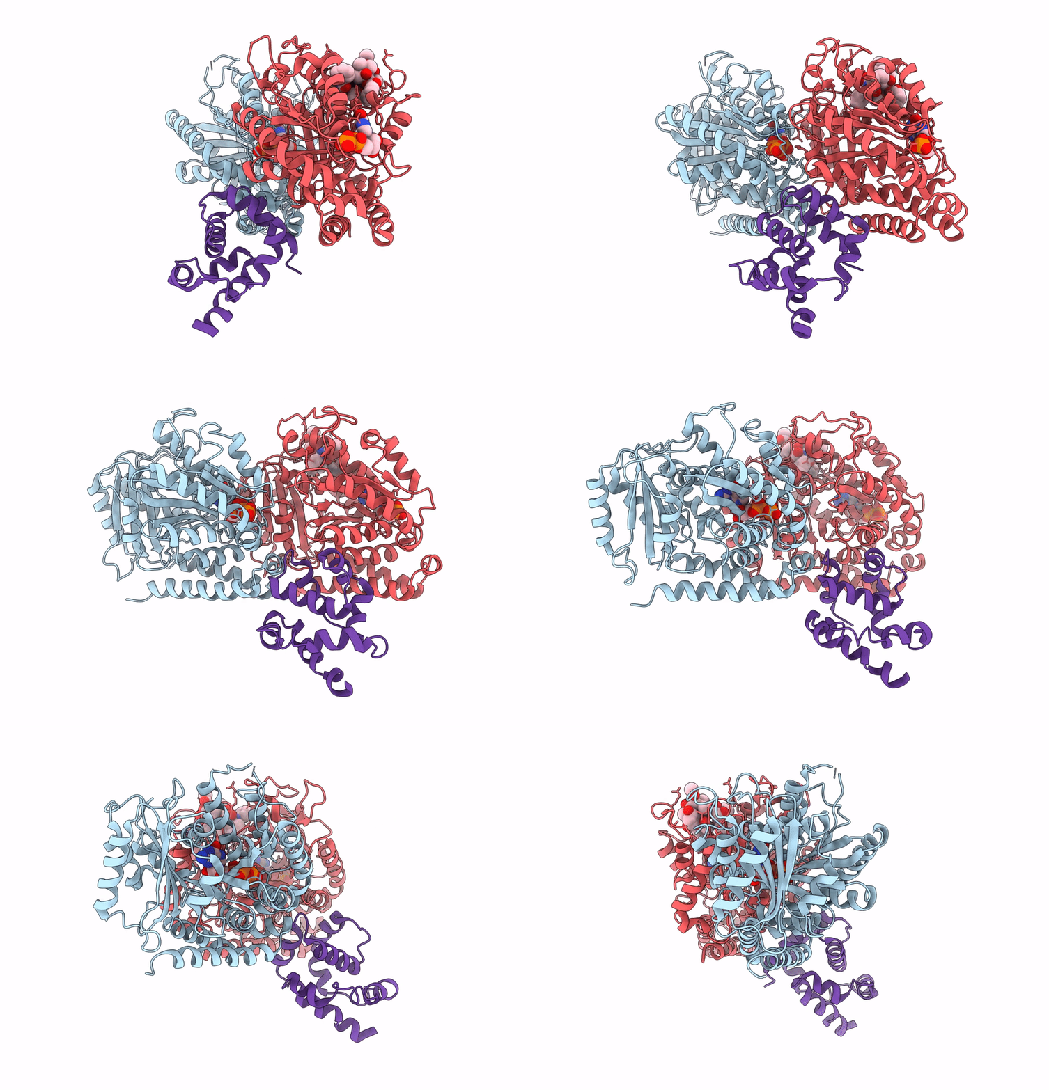
3J6P image
Entry Detail
PDB ID:
3J6P
Title:
Pseudo-atomic model of dynein microtubule binding domain-tubulin complex based on a cryoEM map
Biological Source:
Host Organism:
PDB Version:
Deposition Date:
2014-03-20
Release Date:
2014-12-31
Method Details:
Experimental Method:
Resolution:
8.20 Å
Aggregation State:
FILAMENT
Reconstruction Method:
HELICAL
Macromolecular Entities
Polymer Type:polypeptide(L)
Description:Tubulin alpha-1A chain
Chain IDs:B (auth: A)
Chain Length:451
Number of Molecules:1
Biological Source:Sus scrofa
Polymer Type:polypeptide(L)
Description:Tubulin beta chain
Chain IDs:C (auth: B)
Chain Length:445
Number of Molecules:1
Biological Source:Sus scrofa
Polymer Type:polypeptide(L)
Description:Dynein heavy chain, cytoplasmic
Chain IDs:A (auth: D)
Chain Length:108
Number of Molecules:1
Biological Source:Dictyostelium discoideum
Primary Citation
A flipped ion pair at the dynein-microtubule interface is critical for dynein motility and ATPase activation
J.Cell Biol. 208 211 222 (2015)
PMID: 25583999 DOI: 10.1083/jcb.201407039

Abstact

Dynein is a motor protein that moves on microtubules (MTs) using the energy of adenosine triphosphate (ATP) hydrolysis. To understand its motility mechanism, it is crucial to know how the signal of MT binding is transmitted to the ATPase domain to enhance ATP hydrolysis. However, the molecular basis of signal transmission at the dynein-MT interface remains unclear. Scanning mutagenesis of tubulin identified two residues in α-tubulin, R403 and E416, that are critical for ATPase activation and directional movement of dynein. Electron cryomicroscopy and biochemical analyses revealed that these residues form salt bridges with the residues in the dynein MT-binding domain (MTBD) that work in concert to induce registry change in the stalk coiled coil and activate the ATPase. The R403-E3390 salt bridge functions as a switch for this mechanism because of its reversed charge relative to other residues at the interface. This study unveils the structural basis for coupling between MT binding and ATPase activation and implicates the MTBD in the control of directional movement.

Legend

Protein

Chemical

Disease

Primary Citation of related structures