3J2J image
Deposition Date 2012-10-04
Release Date 2013-07-17
Last Version Date 2024-02-21
Entry Detail
PDB ID:
3J2J
Keywords:
Title:
Empty coxsackievirus A9 capsid
Biological Source:
Source Organism:
Method Details:
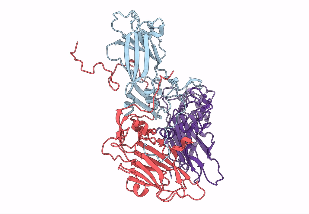
Experimental Method:
Resolution:
9.54 Å
Aggregation State:
PARTICLE
Reconstruction Method:
SINGLE PARTICLE
Macromolecular Entities
Structures with similar UniProt ID
Protein Blast
Polymer Type:polypeptide(L)
Molecule:Protein VP1
Chain IDs:A
Chain Length:222
Number of Molecules:1
Biological Source:Human coxsackievirus A9
Structures with similar UniProt ID
Protein Blast
Polymer Type:polypeptide(L)
Molecule:Protein VP3
Chain IDs:B
Chain Length:238
Number of Molecules:1
Biological Source:Human coxsackievirus A9
Structures with similar UniProt ID
Protein Blast
Polymer Type:polypeptide(L)
Molecule:Protein VP2
Chain IDs:C
Chain Length:252
Number of Molecules:1
Biological Source:Human coxsackievirus A9
Ligand Molecules
Primary Citation
Structural and functional analysis of coxsackievirus A9 integrin {alpha}v{beta}6 binding and uncoating.
J.Virol. 87 3943 3951 (2013)
PMID: 23365426 DOI: 10.1128/JVI.02989-12

Abstact

Coxsackievirus A9 (CVA9) is an important pathogen of the Picornaviridae family. It utilizes cellular receptors from the integrin αv family for binding to its host cells prior to entry and genome release. Among the integrins tested, it has the highest affinity for αvβ6, which recognizes the arginine-glycine-aspartic acid (RGD) loop present on the C terminus of viral capsid protein, VP1. As the atomic model of CVA9 lacks the RGD loop, we used surface plasmon resonance, electron cryo-microscopy, and image reconstruction to characterize the capsid-integrin interactions and the conformational changes on genome release. We show that the integrin binds to the capsid with nanomolar affinity and that the binding of integrin to the virion does not induce uncoating, thereby implying that further steps are required for release of the genome. Electron cryo-tomography and single-particle image reconstruction revealed variation in the number and conformation of the integrins bound to the capsid, with the integrin footprint mapping close to the predicted site for the exposed RGD loop on VP1. Comparison of empty and RNA-filled capsid reconstructions showed that the capsid undergoes conformational changes when the genome is released, so that the RNA-capsid interactions in the N termini of VP1 and VP4 are lost, VP4 is removed, and the capsid becomes more porous, as has been reported for poliovirus 1, human rhinovirus 2, enterovirus 71, and coxsackievirus A7. These results are important for understanding the structural basis of integrin binding to CVA9 and the molecular events leading to CVA9 cell entry and uncoating.

Legend

Protein

Chemical

Disease

Primary Citation of related structures