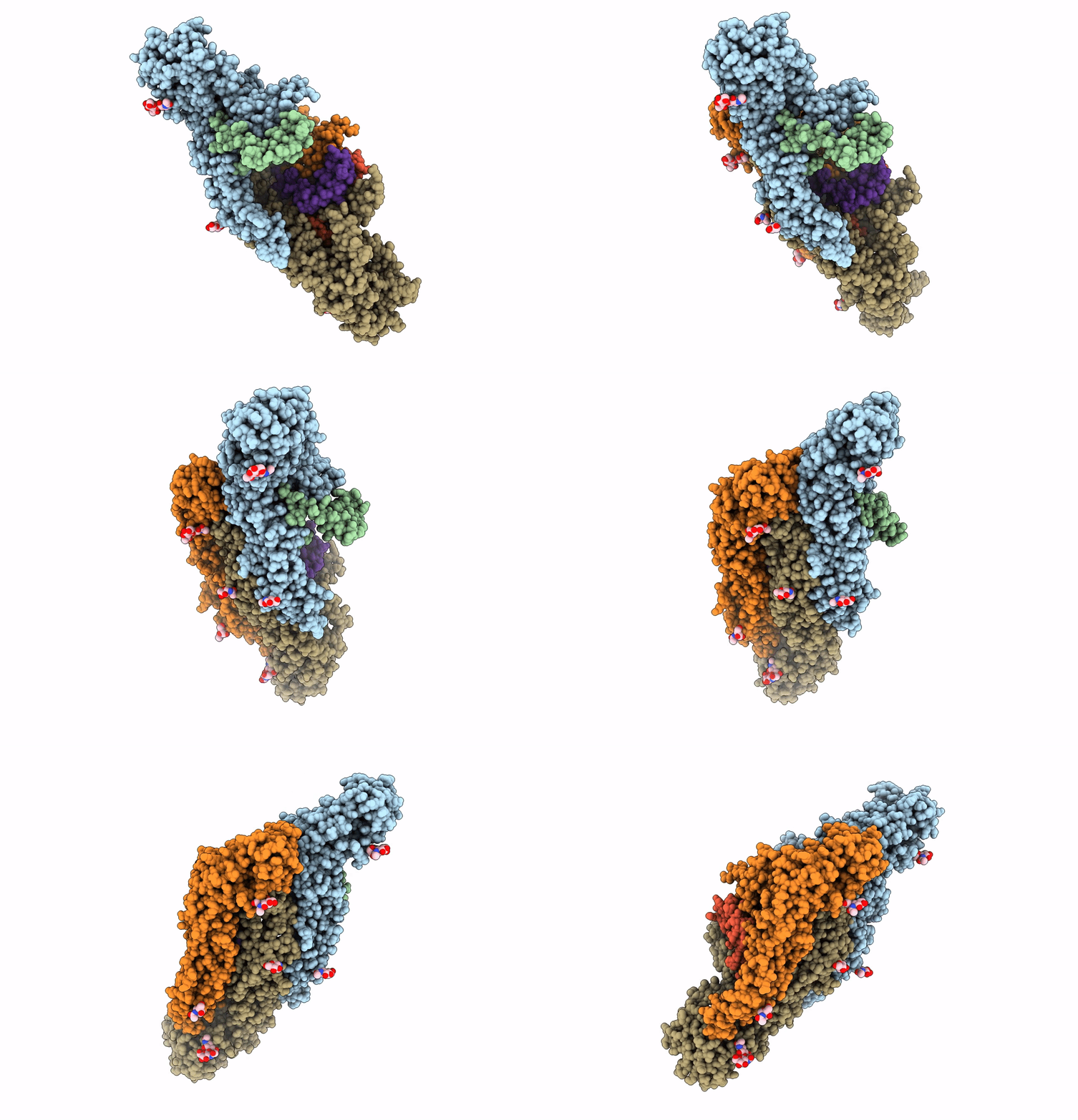
3J27 image
Entry Detail
PDB ID:
3J27
Keywords:
Title:
CryoEM structure of Dengue virus
Biological Source:
Source Organism:
PDB Version:
Deposition Date:
2012-09-26
Release Date:
2012-12-19
Method Details:
Experimental Method:
Resolution:
3.60 Å
Aggregation State:
PARTICLE
Reconstruction Method:
SINGLE PARTICLE
Macromolecular Entities
Polymer Type:polypeptide(L)
Description:Envelope protein E
Chain IDs:A, C, E
Chain Length:495
Number of Molecules:3
Biological Source:Dengue virus 2
Polymer Type:polypeptide(L)
Description:Small envelope protein M
Chain IDs:B, D, F
Chain Length:75
Number of Molecules:3
Biological Source:Dengue virus 2
Modified Residue
Compound ID Chain ID Parent Comp ID Details 2D Image
ASN A ASN GLYCOSYLATION SITE
Ligand Molecules
Primary Citation
Cryo-EM structure of the mature dengue virus at 3.5-A resolution.
Nat.Struct.Mol.Biol. 20 105 110 (2012)
PMID: 23241927 DOI: 10.1038/nsmb.2463

Abstact

Regulated by pH, membrane-anchored proteins E and M function during dengue virus maturation and membrane fusion. Our atomic model of the whole virion from cryo-electron microscopy at 3.5-Å resolution reveals that in the mature virus at neutral extracellular pH, the N-terminal 20-amino-acid segment of M (involving three pH-sensing histidines) latches and thereby prevents spring-loaded E fusion protein from prematurely exposing its fusion peptide. This M latch is fastened at an earlier stage, during maturation at acidic pH in the trans-Golgi network. At a later stage, to initiate infection in response to acidic pH in the late endosome, M releases the latch and exposes the fusion peptide. Thus, M serves as a multistep chaperone of E to control the conformational changes accompanying maturation and infection. These pH-sensitive interactions could serve as targets for drug discovery.

Legend

Protein

Chemical

Disease

Primary Citation of related structures