3IZX image
Deposition Date 2011-01-15
Release Date 2011-06-22
Last Version Date 2024-10-16
Entry Detail
PDB ID:
3IZX
Keywords:
Title:
3.1 Angstrom cryoEM structure of cytoplasmic polyhedrosis virus
Biological Source:
Source Organism:
Method Details:
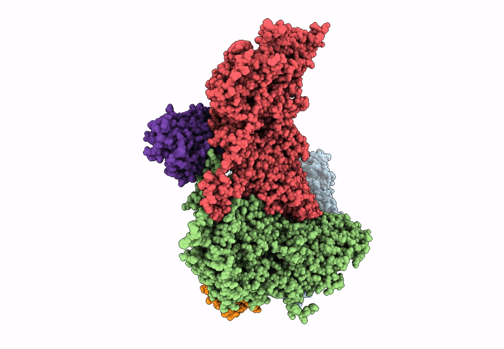
Experimental Method:
Resolution:
3.10 Å
Aggregation State:
PARTICLE
Reconstruction Method:
SINGLE PARTICLE
Macromolecular Entities
Structures with similar UniProt ID
Protein Blast
Polymer Type:polypeptide(L)
Molecule:Structural protein VP3
Chain IDs:A
Chain Length:1058
Number of Molecules:1
Biological Source:Bombyx mori cypovirus 1
Structures with similar UniProt ID
Protein Blast
Polymer Type:polypeptide(L)
Molecule:Capsid protein VP1
Chain IDs:B, C
Chain Length:1333
Number of Molecules:2
Biological Source:Bombyx mori cypovirus 1
Structures with similar UniProt ID
Protein Blast
Polymer Type:polypeptide(L)
Molecule:Viral structural protein 5
Gene (Uniprot):VP5
Chain IDs:D, E
Chain Length:448
Number of Molecules:2
Biological Source:Bombyx mori cypovirus 1
Ligand Molecules
Primary Citation
Atomic Model of CPV Reveals the Mechanism Used by This Single-Shelled Virus to Economically Carry Out Functions Conserved in Multishelled Reoviruses.
Structure 19 652 661 (2011)
PMID: 21565700 DOI: 10.1016/j.str.2011.03.003

Abstact

Unlike the multishelled viruses in the Reoviridae, cytoplasmic polyhedrosis virus (CPV) is single shelled, yet stable and fully capable of carrying out functions conserved within Reoviridae. Here, we report a 3.1 Å resolution cryo electron microscopy structure of CPV and derive its atomic model, consisting of 60 turret proteins (TPs), 120 each of capsid shell proteins (CSPs) and large protrusion proteins (LPPs). Two unique segments of CSP contribute to CPV's stability: an inserted protrusion domain interacting with neighboring proteins, and an N-anchor tying up CSPs together through strong interactions such as β sheet augmentation. Without the need to interact with outer shell proteins, LPP retains only the N-terminal two-third region containing a conserved helix-barrel core and interacts exclusively with CSP. TP is also simplified, containing only domains involved in RNA capping. Our results illustrate how CPV proteins have evolved in a coordinative manner to economically carry out their conserved functions.

Legend

Protein

Chemical

Disease

Primary Citation of related structures
Feedback Form
Name
Email
Institute
Feedback