3IVN image
Deposition Date 2009-09-01
Release Date 2010-01-19
Last Version Date 2023-09-06
Entry Detail
PDB ID:
3IVN
Keywords:
Title:
Structure of the U65C mutant A-riboswitch aptamer from the Bacillus subtilis pbuE operon
Biological Source:
Source Organism:
Host Organism:
Method Details:
Experimental Method:
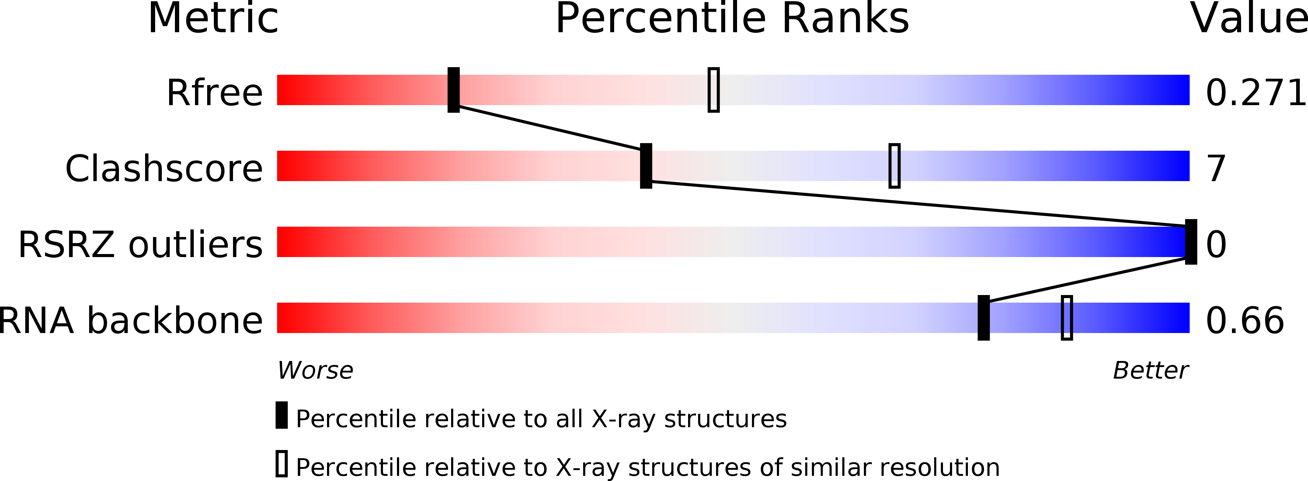
Resolution:
2.80 Å
R-Value Free:
0.27
R-Value Work:
0.22
R-Value Observed:
0.23
Space Group:
C 1 2 1
Macromolecular Entities
Polymer Type:polyribonucleotide
Molecule:A-riboswitch
Mutations:U65C
Chain IDs:A, B
Chain Length:70
Number of Molecules:2
Biological Source:Bacillus subtilis
Primary Citation
Riboswitch structure: an internal residue mimicking the purine ligand.
Nucleic Acids Res. 38 2057 2068 (2010)
PMID: 20022916 DOI: 10.1093/nar/gkp1080

Abstact

The adenine and guanine riboswitches regulate gene expression in response to their purine ligand. X-ray structures of the aptamer moiety of these riboswitches are characterized by a compact fold in which the ligand forms a Watson-Crick base pair with residue 65. Phylogenetic analyses revealed a strict restriction at position 39 of the aptamer that prevents the G39-C65 and A39-U65 combinations, and mutational studies indicate that aptamers with these sequence combinations are impaired for ligand binding. In order to investigate the rationale for sequence conservation at residue 39, structural characterization of the U65C mutant from Bacillus subtilis pbuE adenine riboswitch aptamer was undertaken. NMR spectroscopy and X-ray crystallography studies demonstrate that the U65C mutant adopts a compact ligand-free structure, in which G39 occupies the ligand-binding site of purine riboswitch aptamers. These studies present a remarkable example of a mutant RNA aptamer that adopts a native-like fold by means of ligand mimicking and explain why this mutant is impaired for ligand binding. Furthermore, this work provides a specific insight into how the natural sequence has evolved through selection of nucleotide identities that contribute to formation of the ligand-bound state, but ensures that the ligand-free state remains in an active conformation.

Legend

Protein

Chemical

Disease

Primary Citation of related structures