Deposition Date
2009-08-27
Release Date
2009-10-20
Last Version Date
2023-09-06
Entry Detail
PDB ID:
3IT9
Keywords:
Title:
Crystal structure of Penicillin-Binding Protein 6 (PBP6) from E. coli in apo state
Biological Source:
Source Organism(s):
Escherichia coli (Taxon ID: 562)
Expression System(s):
Method Details:
Experimental Method:
Resolution:
2.10 Å
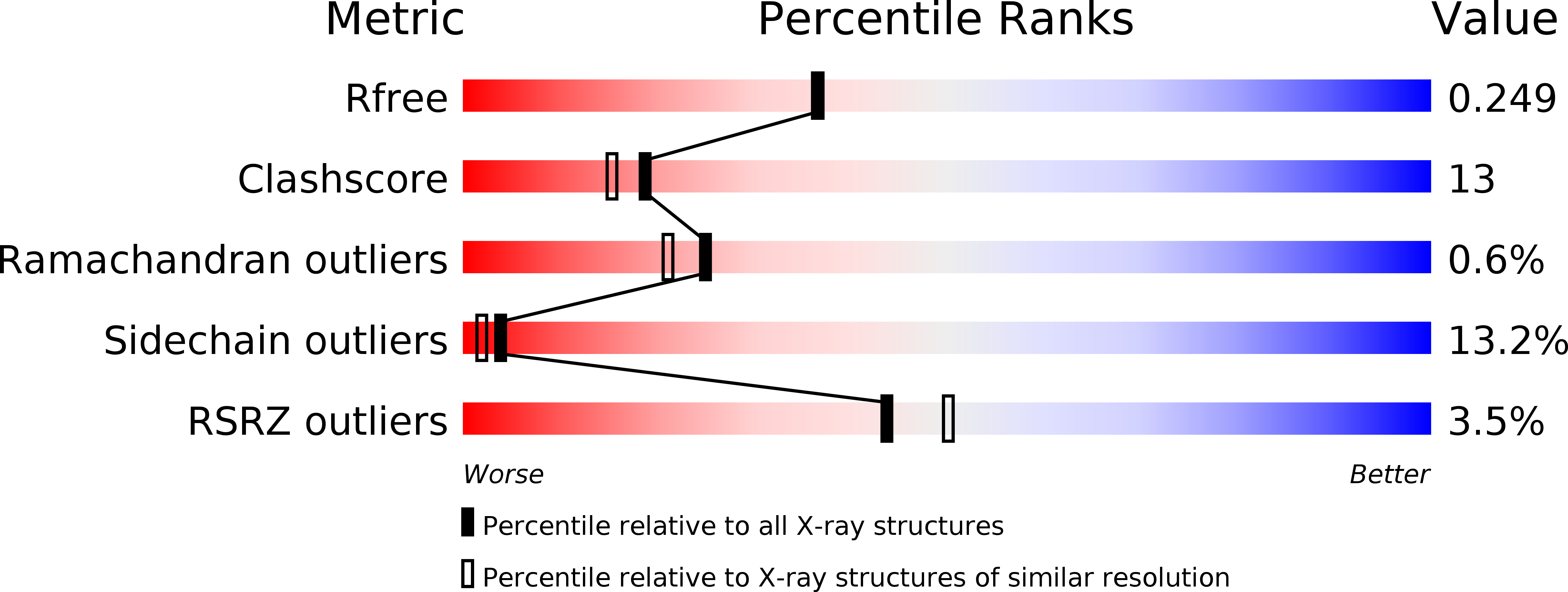
R-Value Free:
0.25
R-Value Work:
0.20
R-Value Observed:
0.21
Space Group:
P 1 21 1