3IT4 image
Deposition Date 2009-08-27
Release Date 2010-03-02
Last Version Date 2023-09-06
Entry Detail
PDB ID:
3IT4
Keywords:
Title:
The Crystal Structure of Ornithine Acetyltransferase from Mycobacterium tuberculosis (Rv1653) at 1.7 A
Biological Source:
Source Organism:
Host Organism:
Method Details:
Experimental Method:
Resolution:
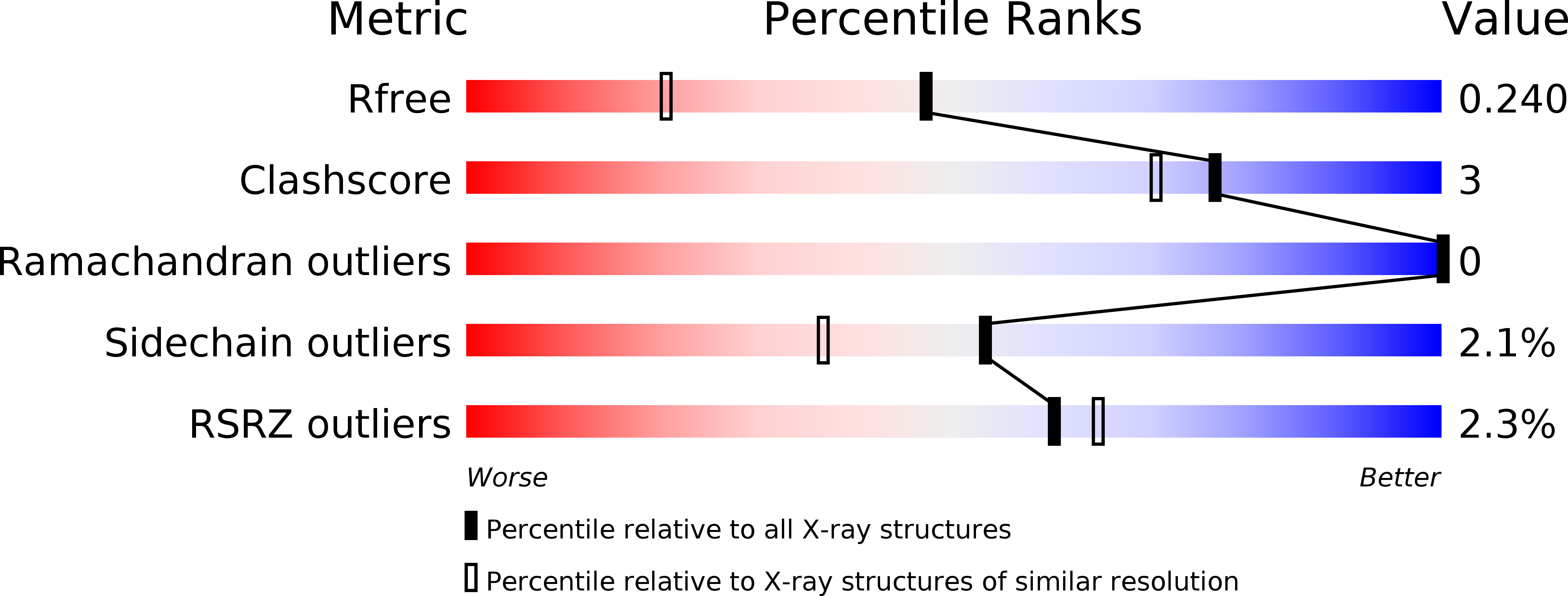
1.70 Å
R-Value Free:
0.24
R-Value Work:
0.20
R-Value Observed:
0.20
Space Group:
P 21 21 21
Macromolecular Entities
Polymer Type:polypeptide(L)
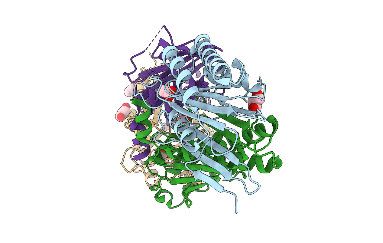
Molecule:Arginine biosynthesis bifunctional protein argJ alpha chain
Chain IDs:A, C
Chain Length:199
Number of Molecules:2
Biological Source:Mycobacterium tuberculosis
Polymer Type:polypeptide(L)
Molecule:Arginine biosynthesis bifunctional protein argJ beta chain
Chain IDs:B, D
Chain Length:205
Number of Molecules:2
Biological Source:Mycobacterium tuberculosis
Primary Citation
The molecular structure of ornithine acetyltransferase from Mycobacterium tuberculosis bound to ornithine, a competitive inhibitor.
J.Mol.Biol. 397 979 990 (2010)
PMID: 20184895 DOI: 10.1016/j.jmb.2010.02.018

Abstact

Mycobacterium tuberculosis ornithine acetyltransferase (Mtb OAT; E.C. 2.3.1.35) is a key enzyme of the acetyl recycling pathway during arginine biosynthesis. It reversibly catalyzes the transfer of the acetyl group from N-acetylornithine (NAORN) to L-glutamate. Mtb OAT is a member of the N-terminal nucleophile fold family of enzymes. The crystal structures of Mtb OAT in native form and in its complex with ornithine (ORN) have been determined at 1.7 and 2.4 A resolutions, respectively. ORN is a competitive inhibitor of this enzyme against L-glutamate as substrate. Although the acyl-enzyme complex of Streptomyces clavuligerus ornithine acetyltransferase has been determined, ours is the first crystal structure to be reported of an ornithine acetyltransferase in complex with an inhibitor. ORN binding does not alter the structure of Mtb OAT globally. However, its presence stabilizes the three C-terminal residues that are disordered and not observed in the native structure. Also, stabilization of the C-terminal residues by ORN reduces the size of the active-site pocket volume in the structure of the ORN complex. The interactions of ORN and the protein residues of Mtb OAT unambiguously delineate the active-site residues of this enzyme in Mtb. Moreover, modeling studies carried out with NAORN based on the structure of the ORN-Mtb OAT complex reveal important interactions of the carbonyl oxygen of the acetyl group of NAORN with the main-chain nitrogen atom of Gly128 and with the side-chain oxygen of Thr127. These interactions likely help in the stabilization of oxyanion formation during enzymatic reaction and also will polarize the carbonyl carbon-oxygen bond, thereby enabling the side-chain atom O(gamma 1) of Thr200 to launch a nucleophilic attack on the carbonyl-carbon atom of the acetyl group of NAORN.

Legend

Protein

Chemical

Disease

Primary Citation of related structures