Deposition Date
2009-08-26
Release Date
2009-09-08
Last Version Date
2023-11-01
Entry Detail
PDB ID:
3ISM
Keywords:
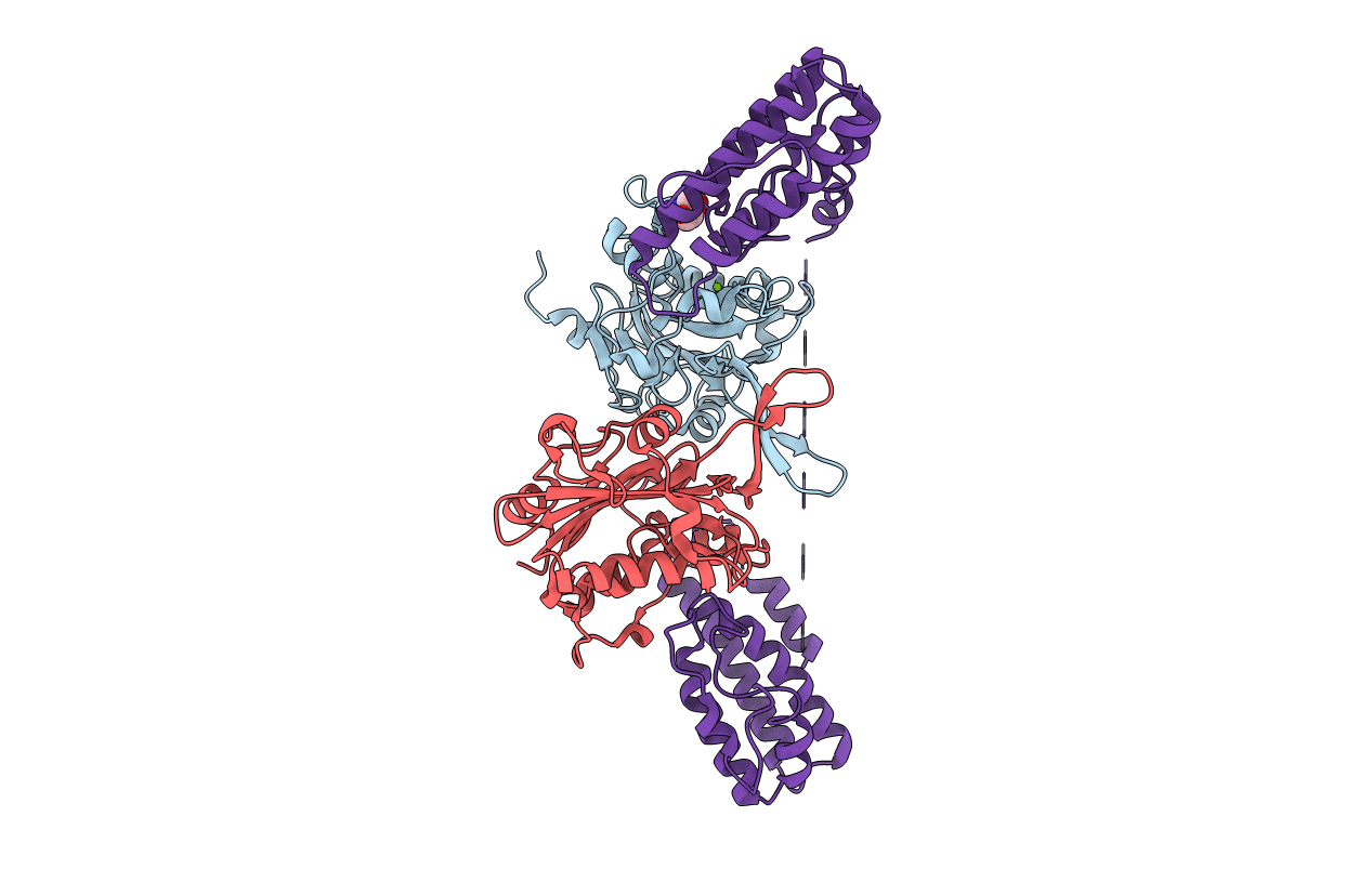
Title:
Crystal structure of the EndoG/EndoGI complex: Mechanism of EndoG inhibition
Biological Source:
Source Organism(s):
Drosophila melanogaster (Taxon ID: 7227)
Expression System(s):
Method Details:
Experimental Method:
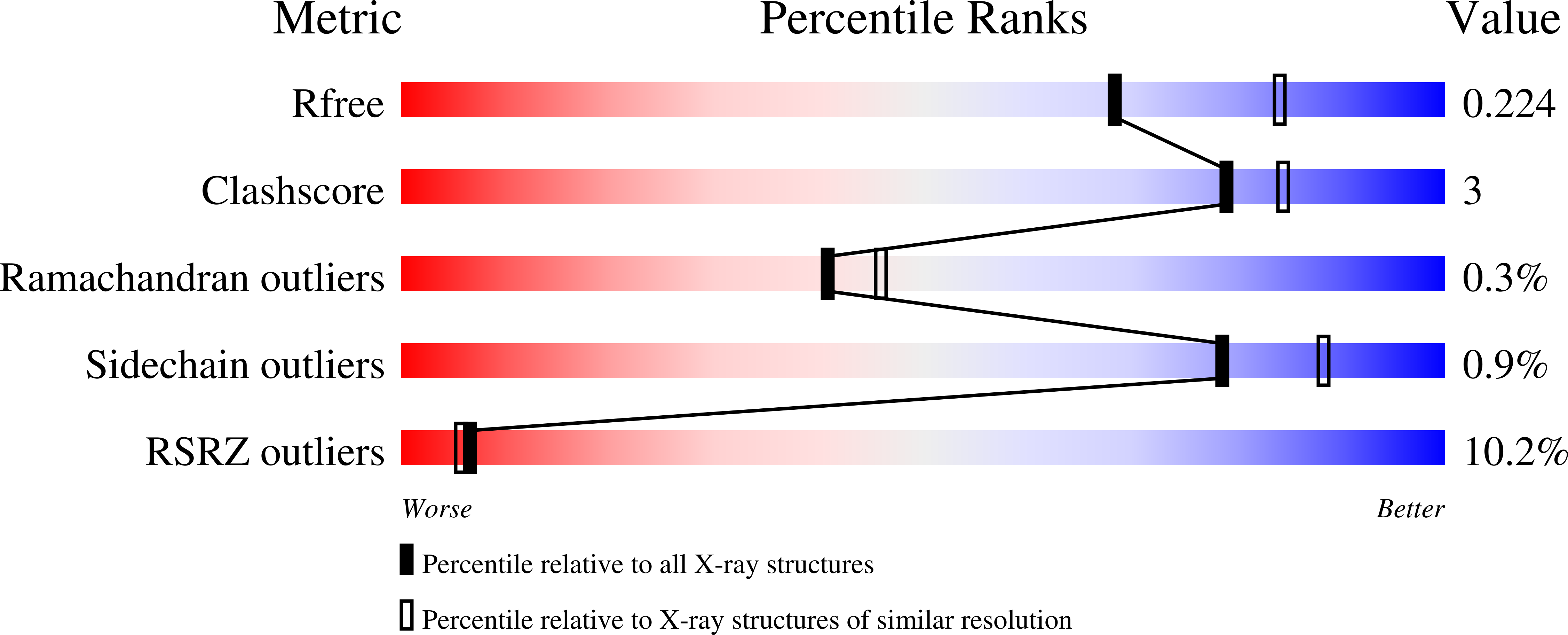
Resolution:
2.20 Å
R-Value Free:
0.22
R-Value Work:
0.18
R-Value Observed:
0.18
Space Group:
P 21 21 21