3IS2 image
Deposition Date 2009-08-25
Release Date 2009-11-03
Last Version Date 2024-10-16
Entry Detail
PDB ID:
3IS2
Title:
2.3 Angstrom Crystal Structure of a Cys71 Sulfenic Acid form of Vivid
Biological Source:
Source Organism:
Host Organism:
Method Details:
Experimental Method:
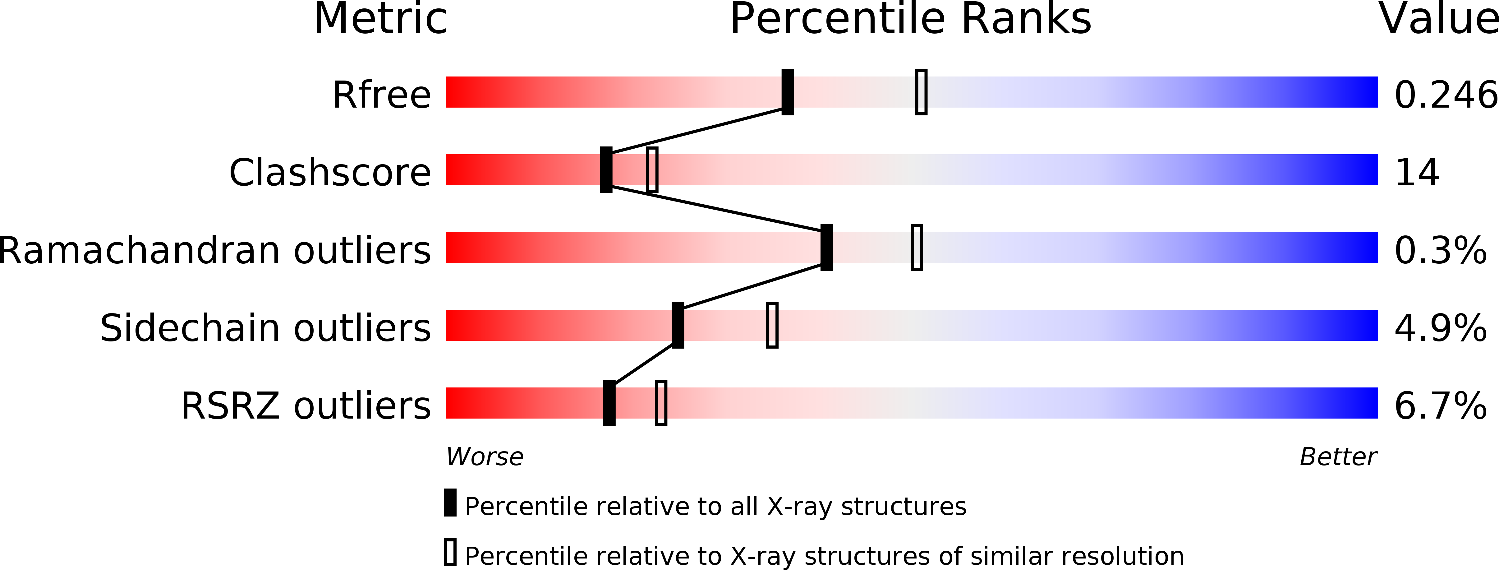
Resolution:
2.30 Å
R-Value Free:
0.25
R-Value Work:
0.22
R-Value Observed:
0.25
Space Group:
P 1 21 1
Macromolecular Entities
Polymer Type:polypeptide(L)
Molecule:Vivid PAS protein VVD
Gene (Uniprot):vvd
Chain IDs:A
Chain Length:154
Number of Molecules:1
Biological Source:Neurospora crassa
Polymer Type:polypeptide(L)
Molecule:Vivid PAS protein VVD
Gene (Uniprot):vvd
Chain IDs:B
Chain Length:154
Number of Molecules:1
Biological Source:Neurospora crassa
Modified Residue
Compound ID Chain ID Parent Comp ID Details 2D Image
CSO A CYS S-HYDROXYCYSTEINE
Ligand Molecules
Primary Citation
Illuminating solution responses of a LOV domain protein with photocoupled small-angle X-ray scattering.
J.Mol.Biol. 393 909 919 (2009)
PMID: 19712683 DOI: 10.1016/j.jmb.2009.08.045

Abstact

The PAS-LOV domain is a signal-transducing component found in a large variety of proteins that is responsible for sensing different stimuli such as light, oxygen, and voltage. The LOV protein VVD regulates blue light responses in the filamentous fungi Neurospora crassa. Using photocoupled, time-resolved small-angle X-ray scattering, we extract the solution protein structure in both dark-adapted and light-activated states. Two distinct dark-adapted conformations are detected in the wild-type protein: a compact structure that corresponds to the crystal structure of the dark-state monomer as well as an extended structure that is well modeled by introducing conformational disorder at the N-terminus of the protein. These conformations are accentuated in carefully selected variants, in which a key residue for propagating structural transitions, Cys71, has been mutated or oxidized. Despite different dark-state conformations, all proteins form a common dimer in response to illumination. Taken together, these data support a reaction scheme that describes the mechanism for light-induced dimerization of VVD. Envelope reconstructions of the transient light-state dimer reveal structures that are best described by a parallel arrangement of subunits that have significantly changed conformation compared to the crystal structure.

Legend

Protein

Chemical

Disease

Primary Citation of related structures