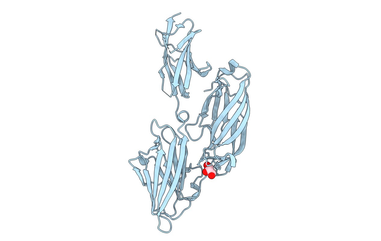
3IS1 image
Entry Detail
PDB ID:
3IS1
Keywords:
Title:
Crystal structure of functional region of UafA from Staphylococcus saprophyticus in C2 form at 2.45 angstrom resolution
Biological Source:
PDB Version:
Deposition Date:
2009-08-24
Release Date:
2010-09-08
Method Details:
Experimental Method:
Resolution:
2.45 Å
R-Value Free:
0.24
R-Value Work:
0.20
R-Value Observed:
0.20
Space Group:
C 1 2 1
Macromolecular Entities
Polymer Type:polypeptide(L)
Description:Uro-adherence factor A
Chain IDs:A (auth: X)
Chain Length:446
Number of Molecules:1
Biological Source:Staphylococcus saprophyticus subsp. saprophyticus
Ligand Molecules
Primary Citation
Crystal structure of the functional region of Uro-adherence factor A from Staphylococcus saprophyticus reveals participation of the B domain in ligand binding
Protein Sci. 20 406 416 (2011)
PMID: 21280131 DOI: 10.1002/pro.573

Abstact

Staphylococci use cell wall-anchored proteins as adhesins to attach to host tissues. Staphylococcus saprophyticus, a uropathogenic species, has a unique cell wall-anchored protein, uro-adherence factor A (UafA), which shows erythrocyte binding activity. To investigate the mechanism of adhesion by UafA, we determined the crystal structure of the functional region of UafA at 1.5 Å resolution. The structure was composed of three domains, designated as the N2, N3, and B domains, arranged in a triangular relative configuration. Hemagglutination inhibition assay with domain-truncated mutants indicated that both N and B domains were necessary for erythrocyte binding. Based on these results, a novel manner of ligand binding in which the B domain acts as a functional domain was proposed as the adhesion mechanism of S. saprophyticus.

Legend

Protein

Chemical

Disease

Primary Citation of related structures