
Deposition Date
2009-08-24
Release Date
2010-09-08
Last Version Date
2023-11-01
Entry Detail
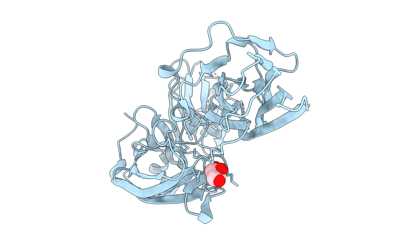
PDB ID:
3IRZ
Keywords:
Title:
Crystal structure of functional region of UafA from Staphylococcus saprophyticus in P212121 form
Biological Source:
Source Organism(s):
Expression System(s):
Method Details:
Experimental Method:
Resolution:
1.70 Å
R-Value Free:
0.21
R-Value Work:
0.19
R-Value Observed:
0.19
Space Group:
P 21 21 21
