3IQD image
Deposition Date 2009-08-20
Release Date 2010-08-25
Last Version Date 2023-11-01
Entry Detail
PDB ID:
3IQD
Keywords:
Title:
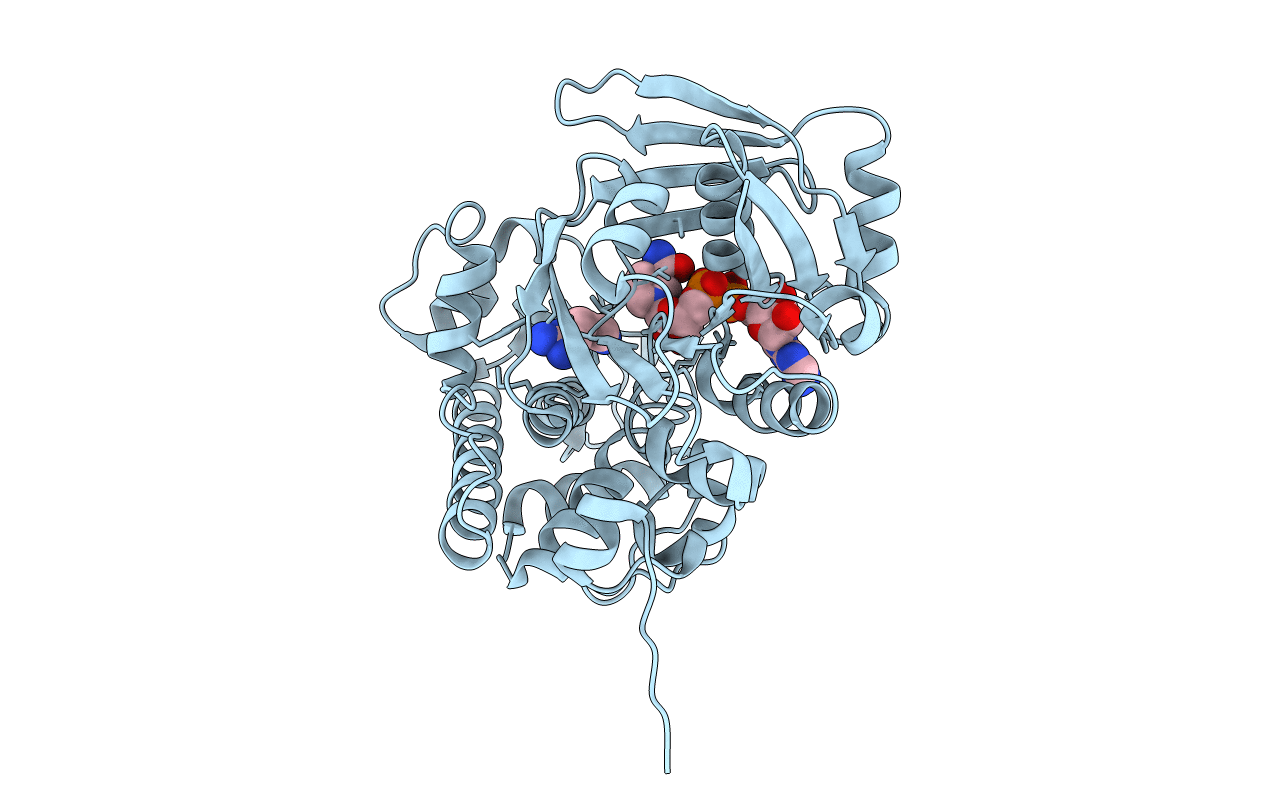
Structure of Octopine-dehydrogenase in complex with NADH and Agmatine
Biological Source:
Source Organism:
Pecten maximus (Taxon ID: 6579)
Host Organism:
Method Details:
Experimental Method:
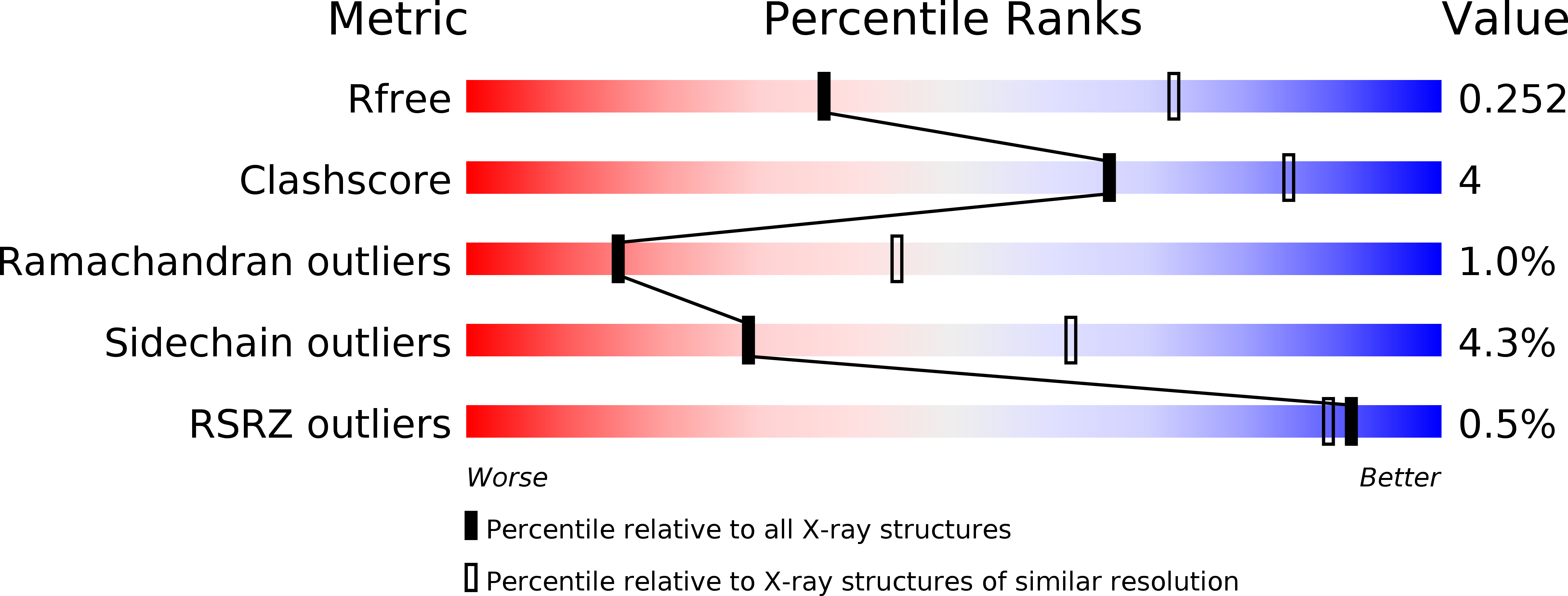
Resolution:
2.80 Å
R-Value Free:
0.25
R-Value Work:
0.20
R-Value Observed:
0.20
Space Group:
P 41 21 2
Macromolecular Entities
Structures with similar UniProt ID
Protein Blast
Polymer Type:polypeptide(L)
Molecule:Octopine dehydrogenase
Gene (Uniprot):odh1
Chain IDs:A (auth: B)
Chain Length:404
Number of Molecules:1
Biological Source:Pecten maximus
Primary Citation
Insights into the mechanism of ligand binding to octopine dehydrogenase from Pecten maximus by NMR and crystallography
Plos One 5 e12312 e12312 (2010)
PMID: 20808820 DOI: 10.1371/journal.pone.0012312

Abstact

Octopine dehydrogenase (OcDH) from the adductor muscle of the great scallop, Pecten maximus, catalyzes the NADH dependent, reductive condensation of L-arginine and pyruvate to octopine, NAD(+), and water during escape swimming and/or subsequent recovery. The structure of OcDH was recently solved and a reaction mechanism was proposed which implied an ordered binding of NADH, L-arginine and finally pyruvate. Here, the order of substrate binding as well as the underlying conformational changes were investigated by NMR confirming the model derived from the crystal structures. Furthermore, the crystal structure of the OcDH/NADH/agmatine complex was determined which suggests a key role of the side chain of L-arginine in protein cataylsis. Thus, the order of substrate binding to OcDH as well as the molecular signals involved in octopine formation can now be described in molecular detail.

Legend

Protein

Chemical

Disease

Primary Citation of related structures
Feedback Form
Name
Email
Institute
Feedback