Deposition Date
2009-08-17
Release Date
2009-12-29
Last Version Date
2023-09-06
Entry Detail
PDB ID:
3IPM
Keywords:
Title:
Crystal Structure of Archaeal 20S Proteasome in Complex with the C-terminus of PAN
Biological Source:
Source Organism(s):
Thermoplasma acidophilum (Taxon ID: 2303)
Trypanosoma brucei brucei (Taxon ID: 5691)
Methanocaldococcus jannaschii (Taxon ID: 2190)
Trypanosoma brucei brucei (Taxon ID: 5691)
Methanocaldococcus jannaschii (Taxon ID: 2190)
Expression System(s):
Method Details:
Experimental Method:
Resolution:
4.00 Å
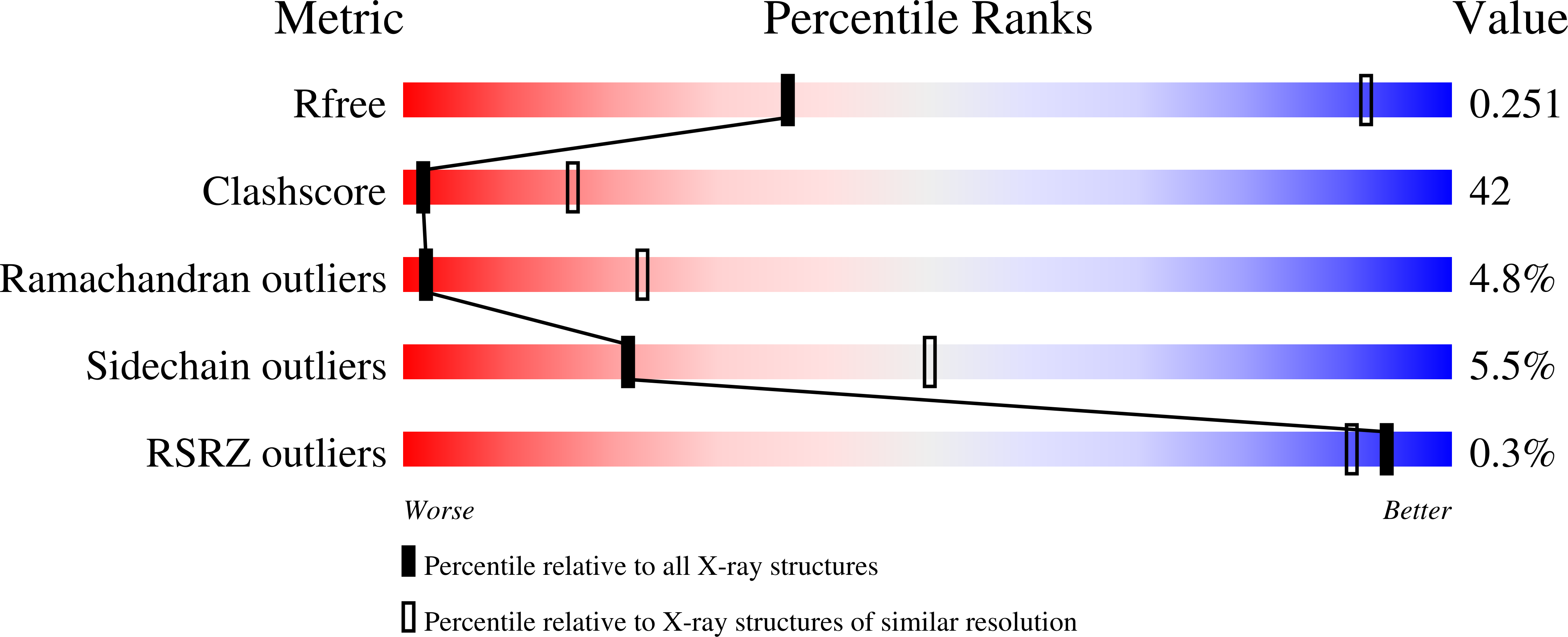
R-Value Free:
0.28
R-Value Work:
0.24
R-Value Observed:
0.25
Space Group:
P 42 2 2