3IPK image
Deposition Date 2009-08-17
Release Date 2010-03-31
Last Version Date 2023-09-06
Entry Detail
PDB ID:
3IPK
Keywords:
Title:
Crystal Structure of A3VP1 of AgI/II of Streptococcus mutans
Biological Source:
Source Organism:
Host Organism:
Method Details:
Experimental Method:
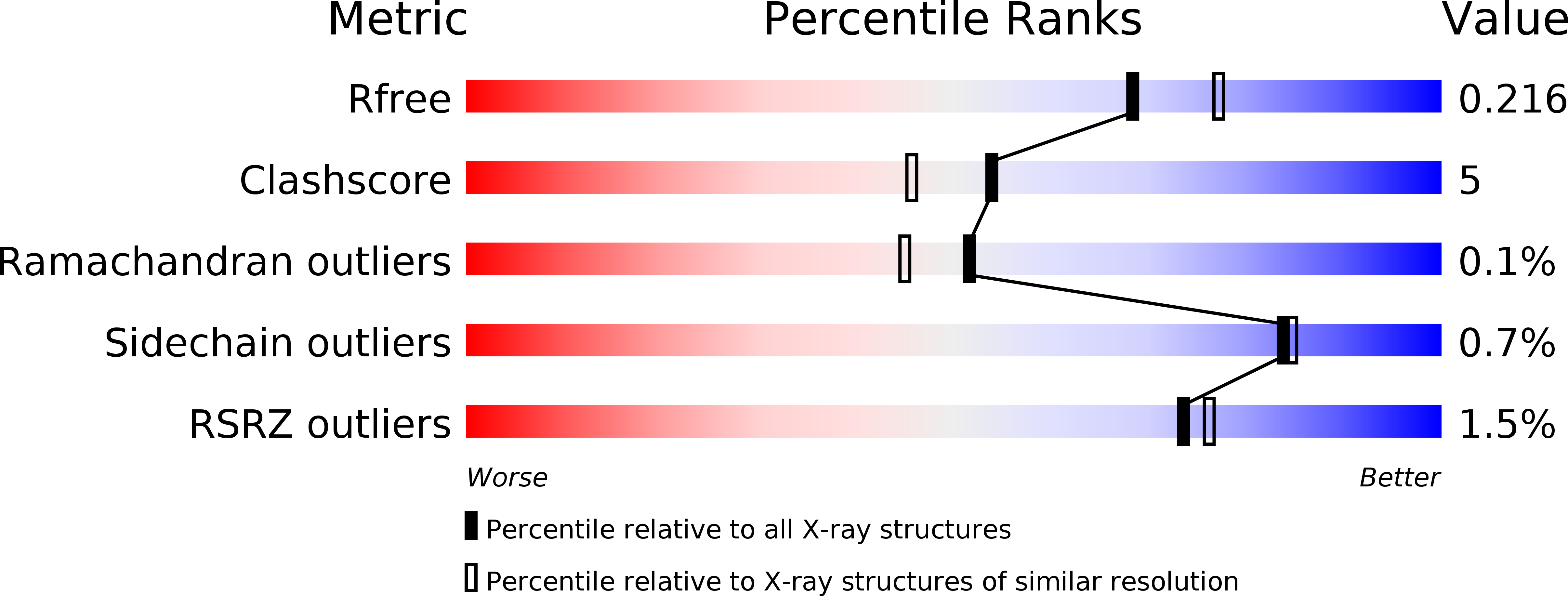
Resolution:
2.04 Å
R-Value Free:
0.22
R-Value Work:
0.18
R-Value Observed:
0.18
Space Group:
P 1 21 1
Macromolecular Entities
Structures with similar UniProt ID
Protein Blast
Polymer Type:polypeptide(L)
Molecule:AgI/II
Chain IDs:A, B
Chain Length:497
Number of Molecules:2
Biological Source:Streptococcus mutans
Primary Citation
Elongated fibrillar structure of a streptococcal adhesin assembled by the high-affinity association of alpha- and PPII-helices.
Proc.Natl.Acad.Sci.USA 107 5983 5988 (2010)
PMID: 20231452 DOI: 10.1073/pnas.0912293107

Abstact

Streptococcus mutans antigen I/II (AgI/II) is a cell surface-localized protein adhesin that interacts with salivary components within the salivary pellicle. AgI/II contributes to virulence and has been studied as an immunological and structural target, but a fundamental understanding of its underlying architecture has been lacking. Here we report a high-resolution (1.8 A) crystal structure of the A(3)VP(1) fragment of S. mutans AgI/II that demonstrates a unique fibrillar form (155 A) through the interaction of two noncontiguous regions in the primary sequence. The A(3) repeat of the alanine-rich domain adopts an extended alpha-helix that intertwines with the P(1) repeat polyproline type II (PPII) helix to form a highly extended stalk-like structure heretofore unseen in prokaryotic or eukaryotic protein structures. Velocity sedimentation studies indicate that full-length AgI/II that contains three A/P repeats extends over 50 nanometers in length. Isothermal titration calorimetry revealed that the high-affinity association between the A(3) and P(1) helices is enthalpically driven. Two distinct binding sites on AgI/II to the host receptor salivary agglutinin (SAG) were identified by surface plasmon resonance (SPR). The current crystal structure reveals that AgI/II family proteins are extended fibrillar structures with the number of alanine- and proline-rich repeats determining their length.

Legend

Protein

Chemical

Disease

Primary Citation of related structures
Feedback Form
Name
Email
Institute
Feedback