3IP5 image
Deposition Date 2009-08-17
Release Date 2010-07-14
Last Version Date 2023-09-06
Entry Detail
PDB ID:
3IP5
Title:
Structure of Atu2422-GABA receptor in complex with alanine
Biological Source:
Source Organism:
Host Organism:
Method Details:
Experimental Method:
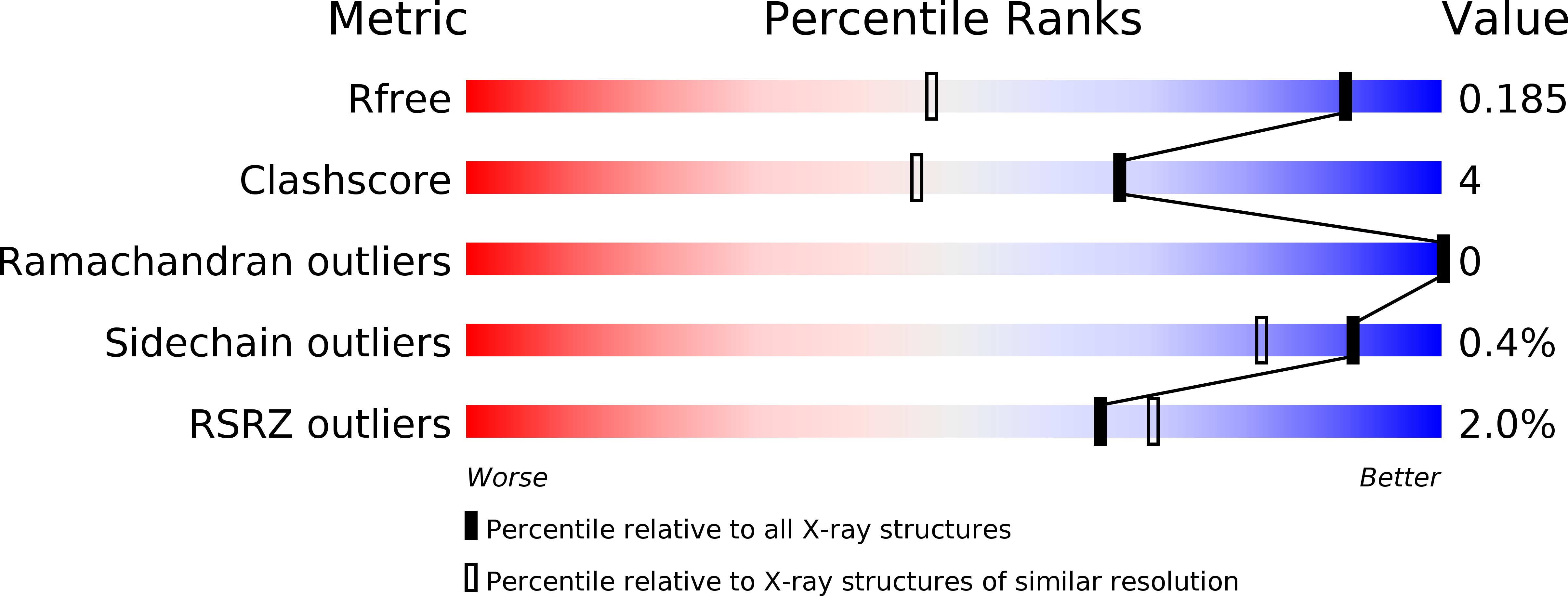
Resolution:
1.35 Å
R-Value Free:
0.18
R-Value Work:
0.16
R-Value Observed:
0.16
Space Group:
C 1 2 1
Macromolecular Entities
Polymer Type:polypeptide(L)
Molecule:ABC transporter, substrate binding protein (Amino acid)
Gene (Uniprot):Atu2422
Chain IDs:A
Chain Length:356
Number of Molecules:1
Biological Source:Agrobacterium tumefaciens
Primary Citation
A conserved mechanism of GABA binding and antagonism is revealed by structure-function analysis of the periplasmic binding protein Atu2422 in Agrobacterium tumefaciens.
J.Biol.Chem. 285 30294 30303 (2010)
PMID: 20630861 DOI: 10.1074/jbc.M110.140715

Abstact

Bacterial periplasmic binding proteins (PBPs) and eukaryotic PBP-like domains (also called as Venus flytrap modules) of G-protein-coupled receptors are involved in extracellular GABA perception. We investigated the structural and functional basis of ligand specificity of the PBP Atu2422, which is implicated in virulence and transport of GABA in the plant pathogen Agrobacterium tumefaciens. Five high-resolution x-ray structures of Atu2422 liganded to GABA, Pro, Ala, and Val and of point mutant Atu2422-F77A liganded to Leu were determined. Structural analysis of the ligand-binding site revealed two essential residues, Phe(77) and Tyr(275), the implication of which in GABA signaling and virulence was confirmed using A. tumefaciens cells expressing corresponding Atu2422 mutants. Phe(77) restricts ligand specificity to α-amino acids with a short lateral chain, which act as antagonists of GABA signaling in A. tumefaciens. Tyr(275) specifically interacts with the GABA γ-amino group. Conservation of these two key residues in proteins phylogenetically related to Atu2422 brought to light a subfamily of PBPs in which all members could bind GABA and short α-amino acids. This work led to the identification of a fingerprint sequence and structural features for defining PBPs that bind GABA and its competitors and revealed their occurrence among host-interacting proteobacteria.

Legend

Protein

Chemical

Disease

Primary Citation of related structures