Deposition Date
2009-08-15
Release Date
2009-12-15
Last Version Date
2023-11-22
Entry Detail
PDB ID:
3IP2
Keywords:
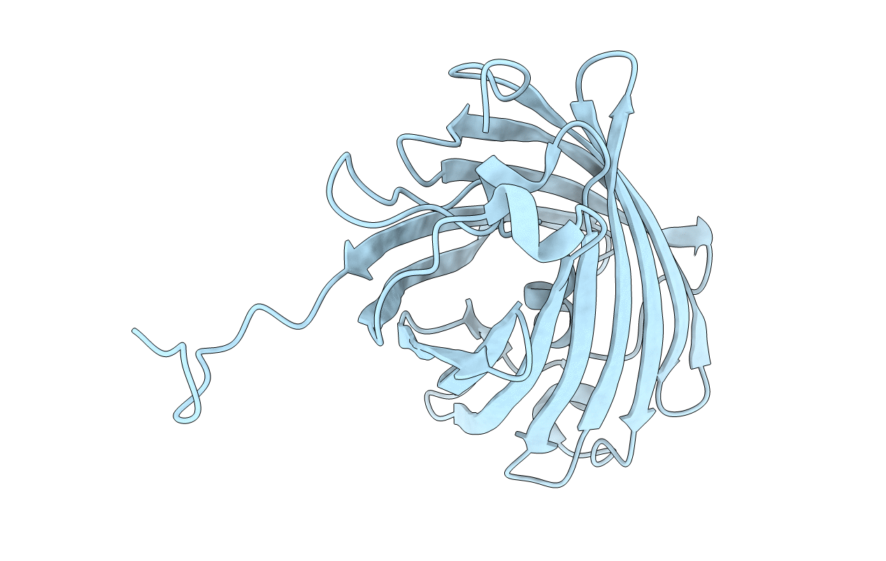
Title:
Crystal structure of red fluorescent protein Neptune at pH 7.0
Biological Source:
Source Organism(s):
Entacmaea quadricolor (Taxon ID: 6118)
Expression System(s):
Method Details:
Experimental Method:
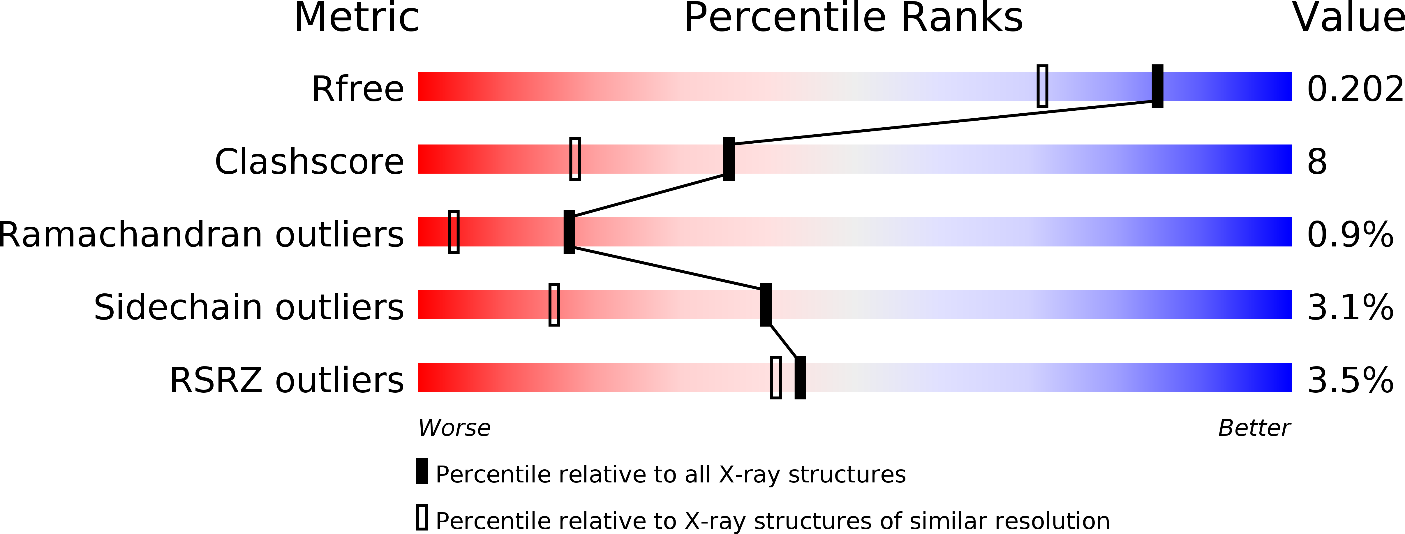
Resolution:
1.60 Å
R-Value Free:
0.20
R-Value Work:
0.17
R-Value Observed:
0.17
Space Group:
P 42 21 2