3IOM image
Entry Detail
PDB ID:
3IOM
Keywords:
Title:
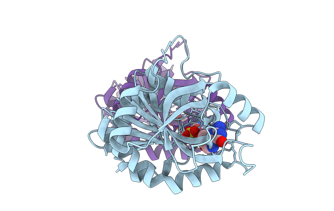
Crystal structure of Purine Nucleoside Phosphorylase from Mycobacterium tuberculosis in complex with 2'-Deoxyguanosine
Biological Source:
Host Organism:
PDB Version:
Deposition Date:
2009-08-14
Release Date:
2011-01-12
Method Details:
Experimental Method:
Resolution:
2.14 Å
R-Value Free:
0.24
R-Value Work:
0.17
R-Value Observed:
0.17
Space Group:
H 3
Macromolecular Entities
Polymer Type:polypeptide(L)
Description:Purine nucleoside phosphorylase
Chain IDs:A, B
Chain Length:268
Number of Molecules:2
Biological Source:Mycobacterium tuberculosis
Primary Citation
Crystallographic and docking studies of purine nucleoside phosphorylase from Mycobacterium tuberculosis.
Bioorg.Med.Chem. 18 4769 4774 (2010)
PMID: 20570524 DOI: 10.1016/j.bmc.2010.05.009

Abstact

This work describes for the first time the structure of purine nucleoside phosphorylase from Mycobacterium tuberculosis (MtPNP) in complex with sulfate and its natural substrate, 2'-deoxyguanosine, and its application to virtual screening. We report docking studies of a set of molecules against this structure. Application of polynomial empirical scoring function was able to rank docking solutions with good predicting power which opens the possibility to apply this new criterion to analyze docking solutions and screen small-molecule databases for new chemical entities to inhibit MtPNP.

Legend

Protein

Chemical

Disease

Primary Citation of related structures