Deposition Date
2009-08-13
Release Date
2009-10-27
Last Version Date
2023-09-06
Entry Detail
PDB ID:
3IO4
Keywords:
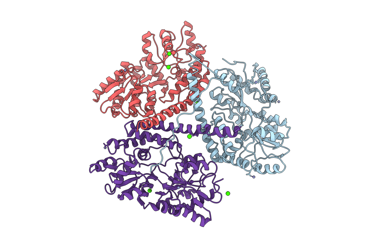
Title:
Huntingtin amino-terminal region with 17 Gln residues - Crystal C90
Biological Source:
Source Organism(s):
Escherichia coli O157:H7 (Taxon ID: 83334)
Homo sapiens (Taxon ID: 9606)
Homo sapiens (Taxon ID: 9606)
Expression System(s):
Method Details:
Experimental Method:
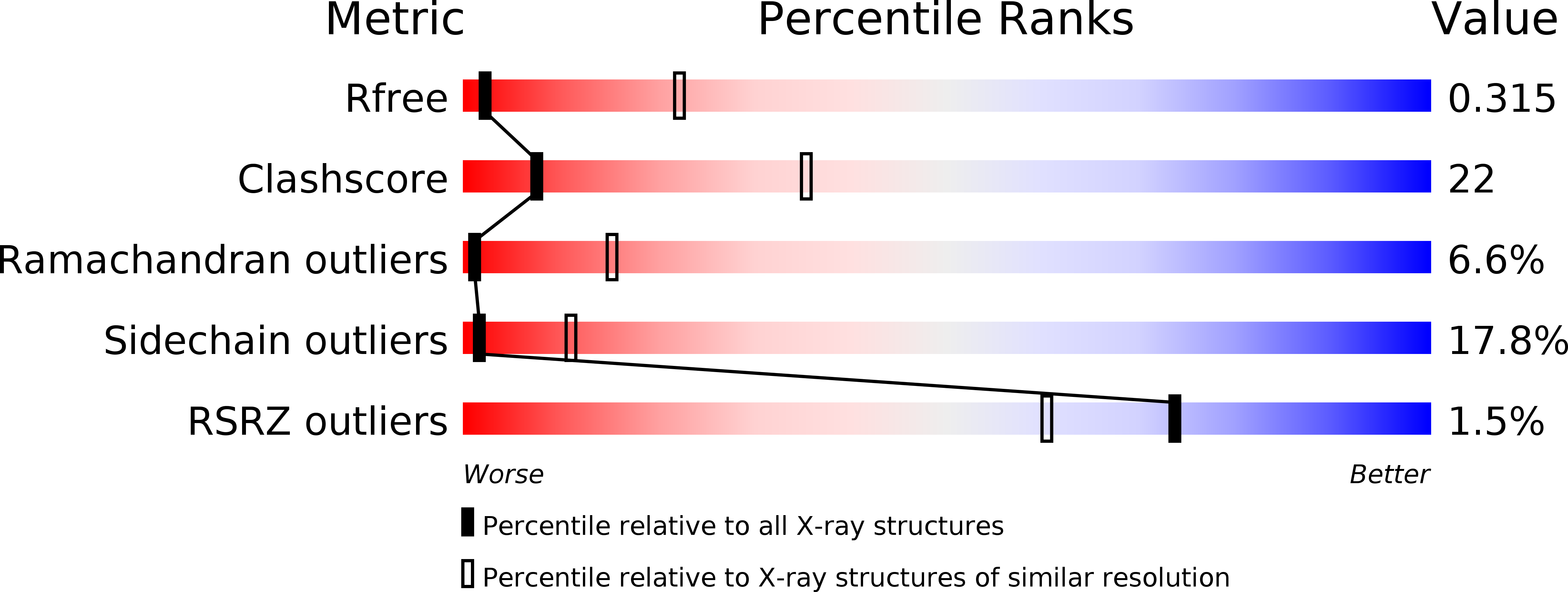
Resolution:
3.63 Å
R-Value Free:
0.30
R-Value Work:
0.25
R-Value Observed:
0.25
Space Group:
C 1 2 1