
Deposition Date
2009-08-11
Release Date
2010-08-04
Last Version Date
2023-11-01
Entry Detail
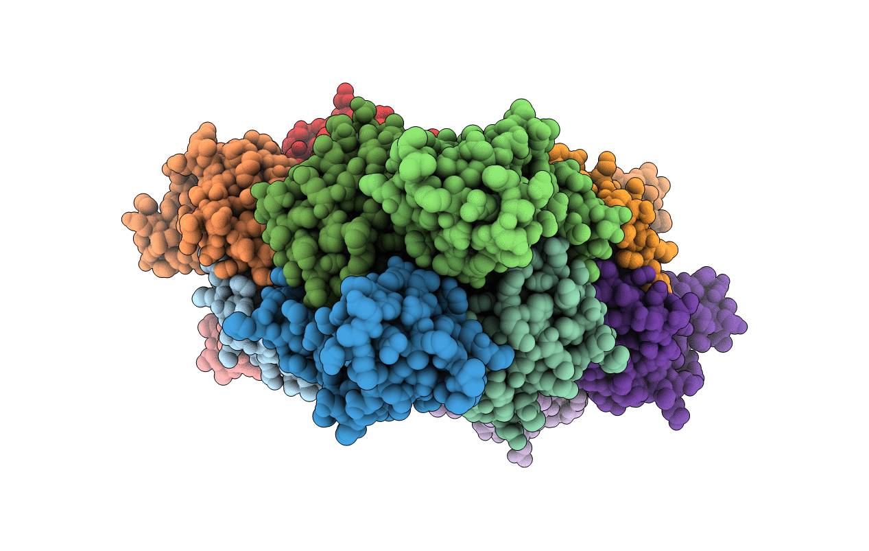
PDB ID:
3IMP
Keywords:
Title:
New crystal form of the C-terminal domain of Helicobacter pylori MotB (residues 125-256)
Biological Source:
Source Organism(s):
Helicobacter pylori (Taxon ID: 85962)
Expression System(s):
Method Details:
Experimental Method:
Resolution:
2.50 Å
R-Value Free:
0.24
R-Value Work:
0.18
R-Value Observed:
0.18
Space Group:
P 1 21 1
