Deposition Date
2009-08-09
Release Date
2010-02-02
Last Version Date
2024-11-20
Entry Detail
PDB ID:
3IM3
Keywords:
Title:
Crystal structure of PKA RI alpha dimerization/docking domain
Biological Source:
Source Organism(s):
Bos taurus (Taxon ID: 9913)
Expression System(s):
Method Details:
Experimental Method:
Resolution:
2.00 Å
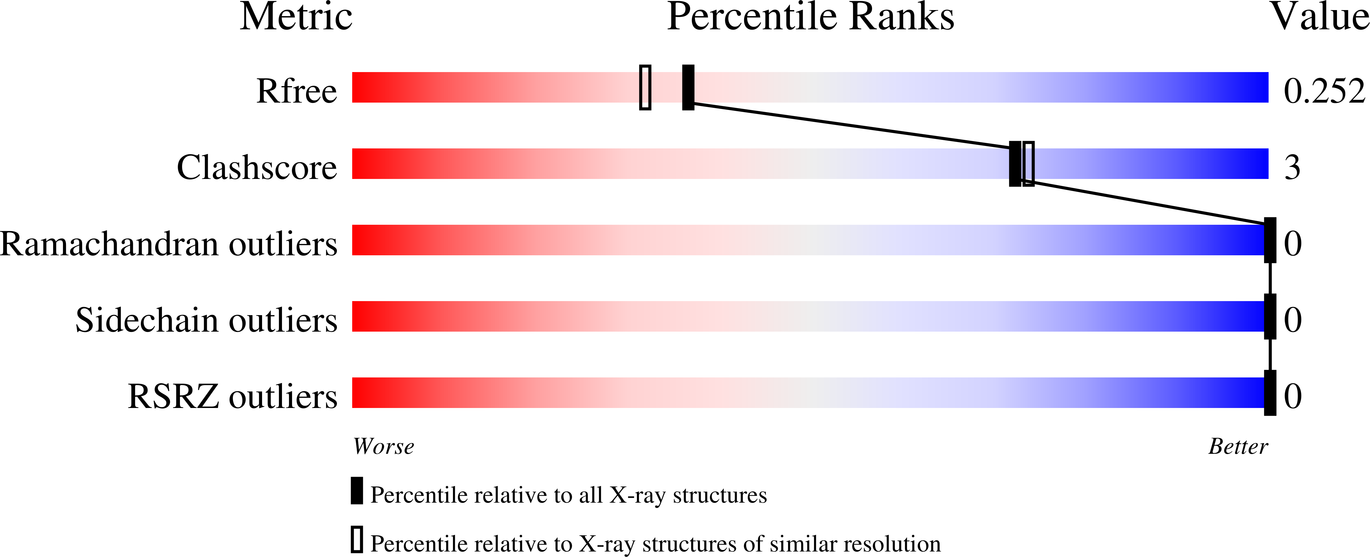
R-Value Free:
0.24
R-Value Work:
0.18
R-Value Observed:
0.19
Space Group:
P 62 2 2