Deposition Date
2009-08-07
Release Date
2010-04-07
Last Version Date
2024-02-21
Entry Detail
Biological Source:
Source Organism(s):
Aspergillus parasiticus (Taxon ID: 5067)
Expression System(s):
Method Details:
Experimental Method:
Resolution:
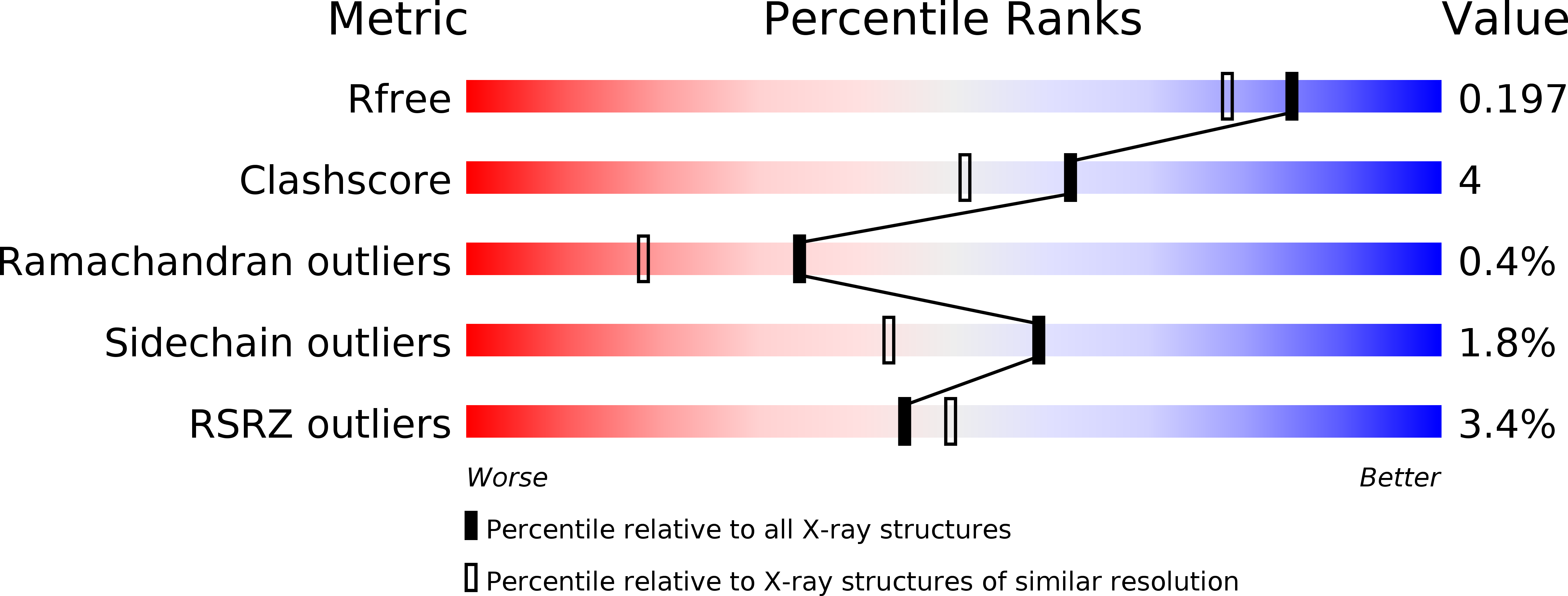
1.70 Å
R-Value Free:
0.20
R-Value Work:
0.17
Space Group:
P 21 21 21