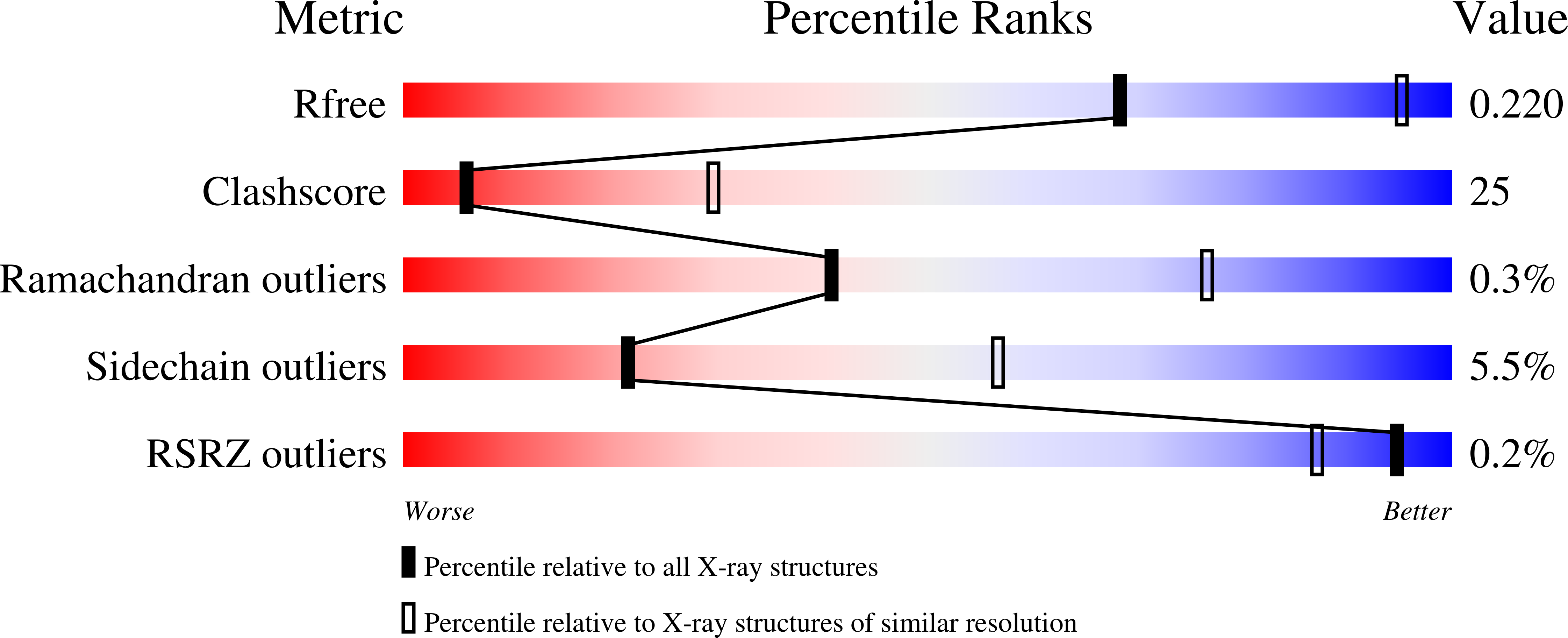
3IL4 image
Deposition Date 2009-08-06
Release Date 2009-08-25
Last Version Date 2024-11-20
Entry Detail
PDB ID:
3IL4
Keywords:
Title:
Structure of E. faecalis FabH in complex with acetyl CoA
Biological Source:
Source Organism:
Host Organism:
Method Details:
Experimental Method:
Resolution:
3.00 Å
R-Value Free:
0.27
R-Value Work:
0.19
R-Value Observed:
0.19
Space Group:
P 1 21 1
Macromolecular Entities
Structures with similar UniProt ID
Protein Blast
Polymer Type:polypeptide(L)
Molecule:3-oxoacyl-[acyl-carrier-protein] synthase 3
Gene (Uniprot):fabH
Mutagens:A192T, N253S
Chain IDs:A, B, C, D
Chain Length:320
Number of Molecules:4
Biological Source:Enterococcus faecalis
Modified Residue
Compound ID Chain ID Parent Comp ID Details 2D Image
CSO A CYS S-HYDROXYCYSTEINE
Ligand Molecules
Primary Citation
Crystal structures of bacterial FabH suggest a molecular basis for the substrate specificity of the enzyme.
Febs Lett. 583 2939 2946 (2009)
PMID: 19665020 DOI: 10.1016/j.febslet.2009.08.001

Abstact

FabH (beta-ketoacyl-acyl carrier protein synthase III) is unique in that it initiates fatty acid biosynthesis, is inhibited by long-chain fatty acids providing means for feedback control of the process, and dictates the fatty acid profile of the organism by virtue of its substrate specificity. We report the crystal structures of bacterial FabH enzymes from four different pathogenic species: Enterococcus faecalis, Haemophilus influenzae, Staphylococcus aureus and Escherichia coli. Structural data on the enzyme from different species show important differences in the architecture of the substrate-binding sites that parallel the inter-species diversity in the substrate specificities of these enzymes.

Legend

Protein

Chemical

Disease

Primary Citation of related structures