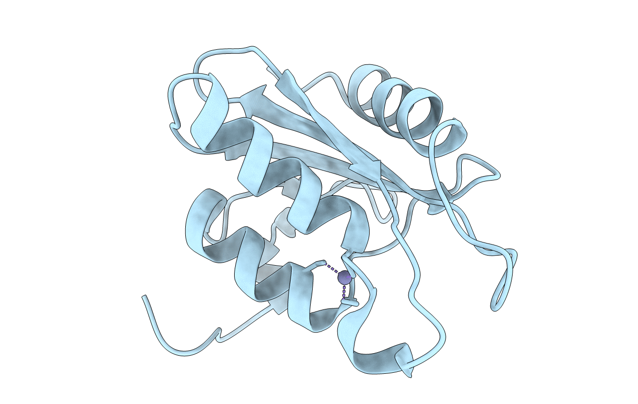
3IJF image
Entry Detail
PDB ID:
3IJF
Keywords:
Title:
Crystal structure of cytidine deaminase from Mycobacterium tuberculosis
Biological Source:
PDB Version:
Deposition Date:
2009-08-04
Release Date:
2010-03-02
Method Details:
Experimental Method:
Resolution:
1.99 Å
R-Value Free:
0.24
R-Value Work:
0.19
R-Value Observed:
0.19
Space Group:
C 2 2 2
Macromolecular Entities
Polymer Type:polypeptide(L)
Description:Cytidine deaminase
Chain IDs:A (auth: X)
Chain Length:133
Number of Molecules:1
Biological Source:Mycobacterium tuberculosis
Ligand Molecules
Primary Citation
Structural and functional analyses of Mycobacterium tuberculosis Rv3315c-encoded metal-dependent homotetrameric cytidine deaminase.
J.Struct.Biol. 169 413 423 (2010)
PMID: 20035876 DOI: 10.1016/j.jsb.2009.12.019

Abstact

The emergence of drug-resistant strains of Mycobacterium tuberculosis, the causative agent of tuberculosis, has exacerbated the treatment and control of this disease. Cytidine deaminase (CDA) is a pyrimidine salvage pathway enzyme that recycles cytidine and 2'-deoxycytidine for uridine and 2'-deoxyuridine synthesis, respectively. A probable M. tuberculosis CDA-coding sequence (cdd, Rv3315c) was cloned, sequenced, expressed in Escherichia coli BL21(DE3), and purified to homogeneity. Mass spectrometry, N-terminal amino acid sequencing, gel filtration chromatography, and metal analysis of M. tuberculosis CDA (MtCDA) were carried out. These results and multiple sequence alignment demonstrate that MtCDA is a homotetrameric Zn(2+)-dependent metalloenzyme. Steady-state kinetic measurements yielded the following parameters: K(m)=1004 microM and k(cat)=4.8s(-1) for cytidine, and K(m)=1059 microM and k(cat)=3.5s(-1) for 2'-deoxycytidine. The pH dependence of k(cat) and k(cat)/K(M) for cytidine indicate that protonation of a single ionizable group with apparent pK(a) value of 4.3 abolishes activity, and protonation of a group with pK(a) value of 4.7 reduces binding. MtCDA was crystallized and crystal diffracted at 2.0 A resolution. Analysis of the crystallographic structure indicated the presence of a Zn(2+) coordinated by three conserved cysteines and the structure exhibits the canonical cytidine deaminase fold.

Legend

Protein

Chemical

Disease

Primary Citation of related structures