Deposition Date
2009-08-03
Release Date
2009-12-08
Last Version Date
2024-10-30
Entry Detail
PDB ID:
3IIR
Keywords:
Title:
Crystal Structure of Miraculin like protein from seeds of Murraya koenigii
Biological Source:
Source Organism(s):
Murraya koenigii (Taxon ID: 311449)
Method Details:
Experimental Method:
Resolution:
2.90 Å
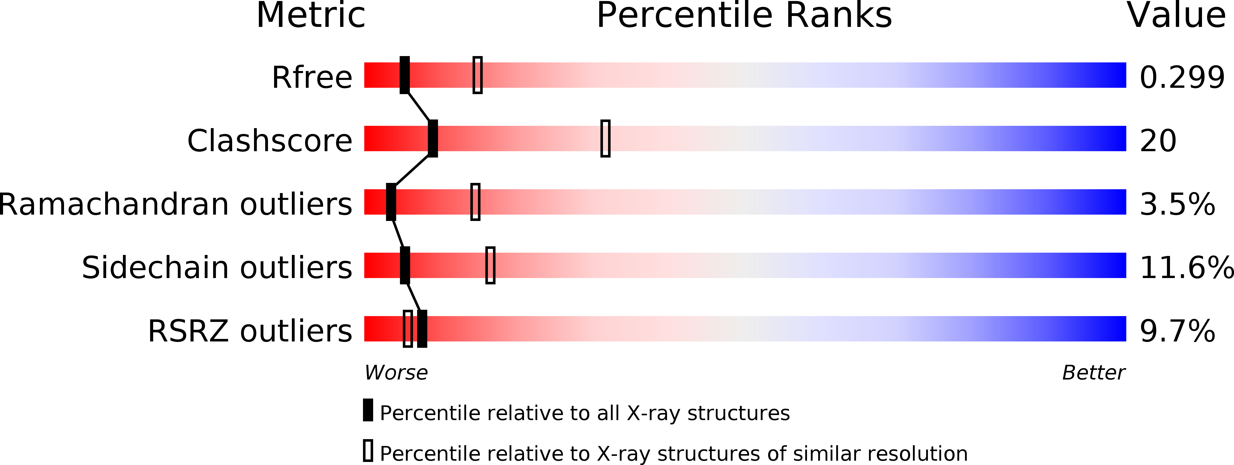
R-Value Free:
0.29
R-Value Work:
0.21
R-Value Observed:
0.21
Space Group:
P 43 21 2