3IG3 image
Deposition Date 2009-07-27
Release Date 2009-08-25
Last Version Date 2024-02-21
Entry Detail
PDB ID:
3IG3
Title:
Crystal structure of mouse Plexin A3 intracellular domain
Biological Source:
Source Organism:
Mus musculus (Taxon ID: 10090)
Host Organism:
Method Details:
Experimental Method:
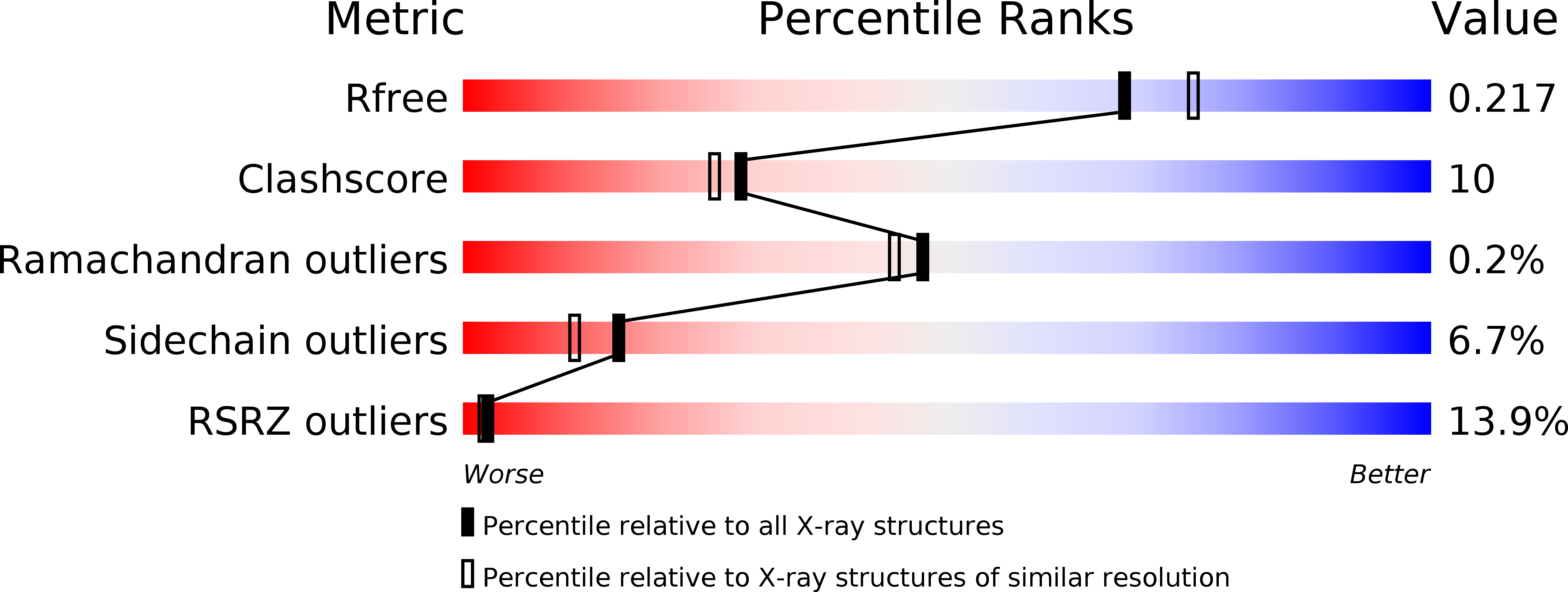
Resolution:
1.99 Å
R-Value Free:
0.22
R-Value Work:
0.18
R-Value Observed:
0.19
Space Group:
C 1 2 1
Macromolecular Entities
Structures with similar UniProt ID
Protein Blast
Polymer Type:polypeptide(L)
Molecule:Plxna3 protein
Chain IDs:A
Chain Length:627
Number of Molecules:1
Biological Source:Mus musculus
Ligand Molecules
Primary Citation
Crystal structure of the plexin A3 intracellular region reveals an autoinhibited conformation through active site sequestration.
Proc.Natl.Acad.Sci.USA 106 15610 15615 (2009)
PMID: 19717441 DOI: 10.1073/pnas.0906923106

Abstact

Plexin cell surface receptors bind to semaphorin ligands and transduce signals for regulating neuronal axon guidance. The intracellular region of plexins is essential for signaling and contains a R-Ras/M-Ras GTPase activating protein (GAP) domain that is divided into two segments by a Rho GTPase-binding domain (RBD). The regulation mechanisms for plexin remain elusive, although it is known that activation requires both binding of semaphorin to the extracellular region and a Rho-family GTPase (Rac1 or Rnd1) to the RBD. Here we report the crystal structure of the plexin A3 intracellular region. The structure shows that the N- and C-terminal portions of the GAP homologous regions together form a GAP domain with an overall fold similar to other Ras GAPs. However, the plexin GAP domain adopts a closed conformation and cannot accommodate R-Ras/M-Ras in its substrate-binding site, providing a structural basis for the autoinhibited state of plexins. A comparison with the plexin B1 RBD/Rnd1 complex structure suggests that Rnd1 binding alone does not induce a conformational change in plexin, explaining the requirement of both semaphorin and a Rho GTPase for activation. The structure also identifies an N-terminal segment that is important for regulation. Both the N-terminal segment and the RBD make extensive interactions with the GAP domain, suggesting the presence of an allosteric network connecting these three domains that integrates semaphorin and Rho GTPase signals to activate the GAP. The importance of these interactions in plexin signaling is shown by both cell-based and in vivo axon guidance assays.

Legend

Protein

Chemical

Disease

Primary Citation of related structures