3IG1 image
Deposition Date 2009-07-27
Release Date 2010-01-26
Last Version Date 2023-09-06
Entry Detail
PDB ID:
3IG1
Keywords:
Title:
HIV-1 Reverse Transcriptase with the Inhibitor beta-Thujaplicinol Bound at the RNase H Active Site
Biological Source:
Source Organism:
Host Organism:
Method Details:
Experimental Method:
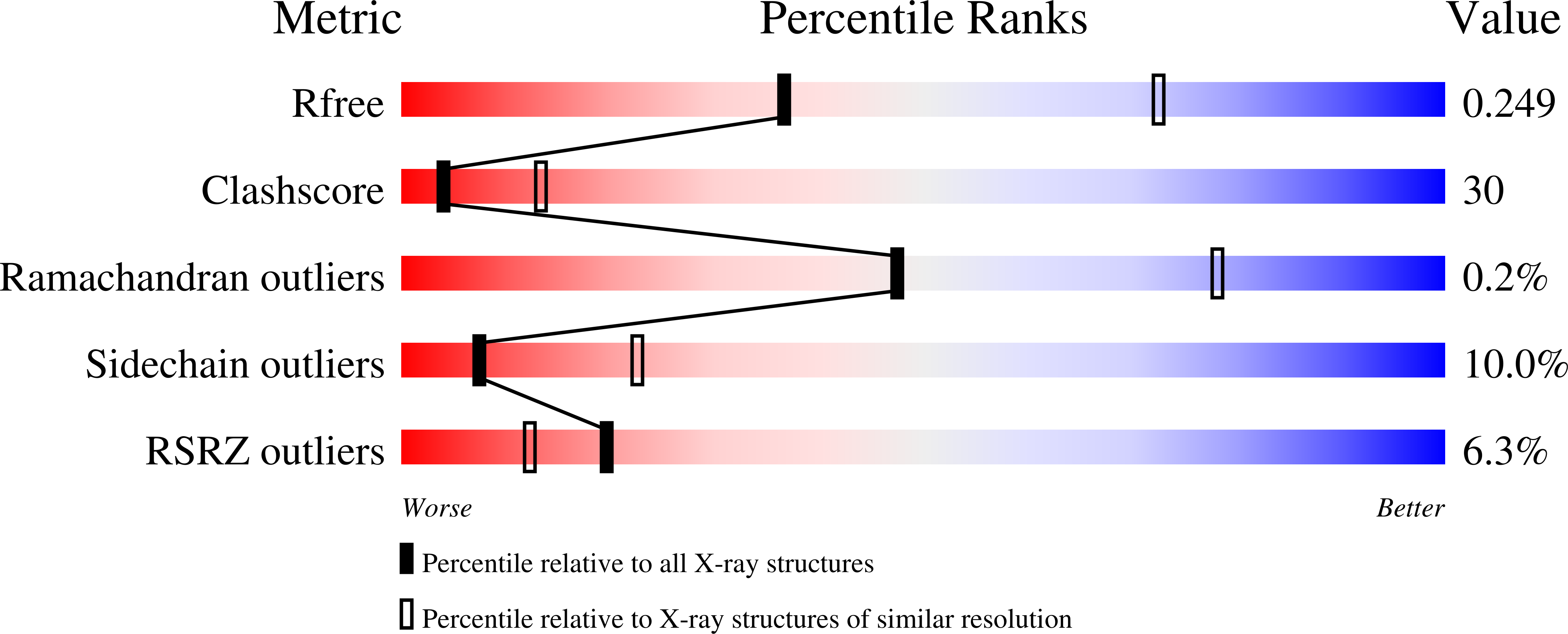
Resolution:
2.80 Å
R-Value Free:
0.25
R-Value Work:
0.23
R-Value Observed:
0.23
Space Group:
C 1 2 1
Macromolecular Entities
Polymer Type:polypeptide(L)
Molecule:HIV-1 Reverse Transcriptase p66 subunit
Gene (Uniprot):gag-pol
Mutations:F759S, C879S
Chain IDs:A
Chain Length:555
Number of Molecules:1
Biological Source:Human immunodeficiency virus type 1 BH10
Polymer Type:polypeptide(L)
Molecule:HIV-1 Reverse Transcriptase p51 subunit
Gene (Uniprot):gag-pol
Mutations:C879S
Chain IDs:B
Chain Length:428
Number of Molecules:1
Biological Source:Human immunodeficiency virus type 1 BH10
Primary Citation
Structure of HIV-1 reverse transcriptase with the inhibitor beta-Thujaplicinol bound at the RNase H active site.
Structure 17 1625 1635 (2009)
PMID: 20004166 DOI: 10.1016/j.str.2009.09.016

Abstact

Novel inhibitors are needed to counteract the rapid emergence of drug-resistant HIV variants. HIV-1 reverse transcriptase (RT) has both DNA polymerase and RNase H (RNH) enzymatic activities, but approved drugs that inhibit RT target the polymerase. Inhibitors that act against new targets, such as RNH, should be effective against all of the current drug-resistant variants. Here, we present 2.80 A and 2.04 A resolution crystal structures of an RNH inhibitor, beta-thujaplicinol, bound at the RNH active site of both HIV-1 RT and an isolated RNH domain. beta-thujaplicinol chelates two divalent metal ions at the RNH active site. We provide biochemical evidence that beta-thujaplicinol is a slow-binding RNH inhibitor with noncompetitive kinetics and suggest that it forms a tropylium ion that interacts favorably with RT and the RNA:DNA substrate.

Legend

Protein

Chemical

Disease

Primary Citation of related structures