Deposition Date
2009-07-26
Release Date
2010-06-16
Last Version Date
2023-11-15
Entry Detail
PDB ID:
3IFW
Keywords:
Title:
Crystal structure of the S18Y variant of ubiquitin carboxy terminal hydrolase L1 bound to ubiquitin vinylmethylester.
Biological Source:
Source Organism(s):
Homo sapiens (Taxon ID: 9606)
Expression System(s):
Method Details:
Experimental Method:
Resolution:
2.40 Å
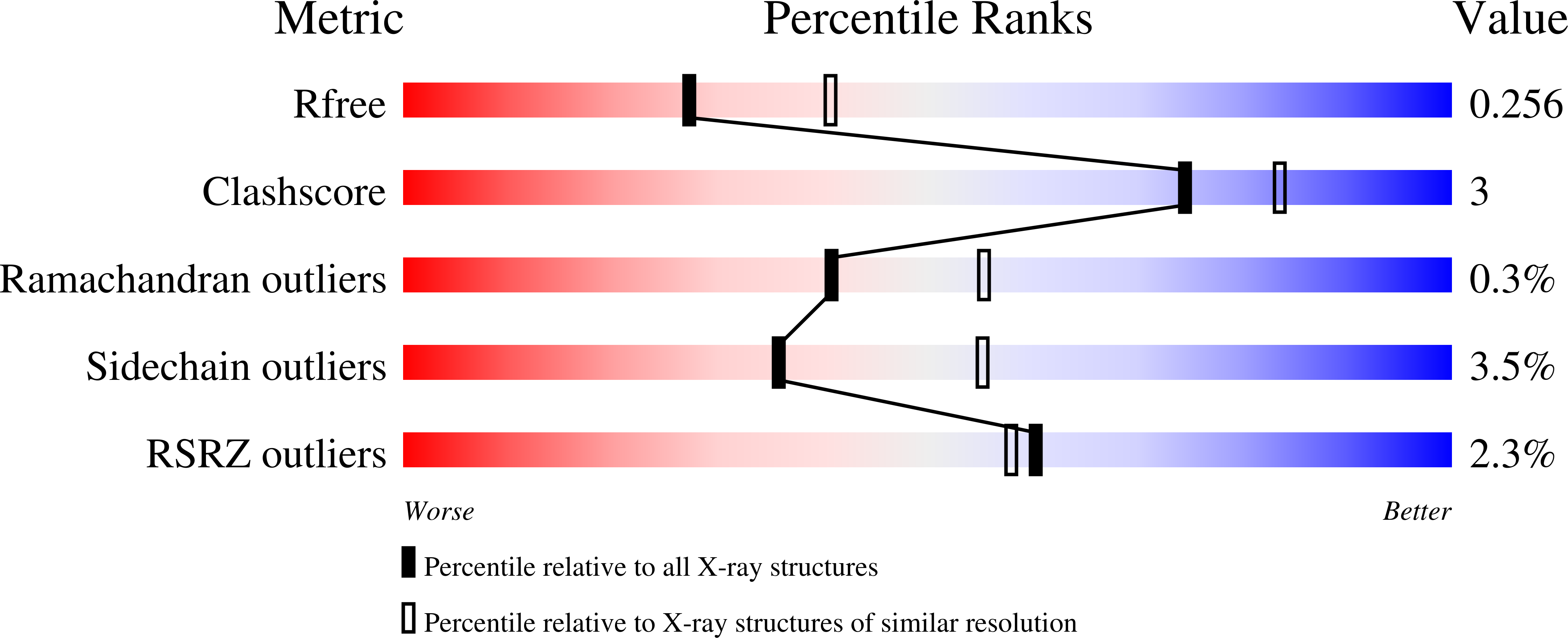
R-Value Free:
0.25
R-Value Work:
0.20
R-Value Observed:
0.21
Space Group:
H 3 2