3IFC image
Entry Detail
PDB ID:
3IFC
Keywords:
Title:
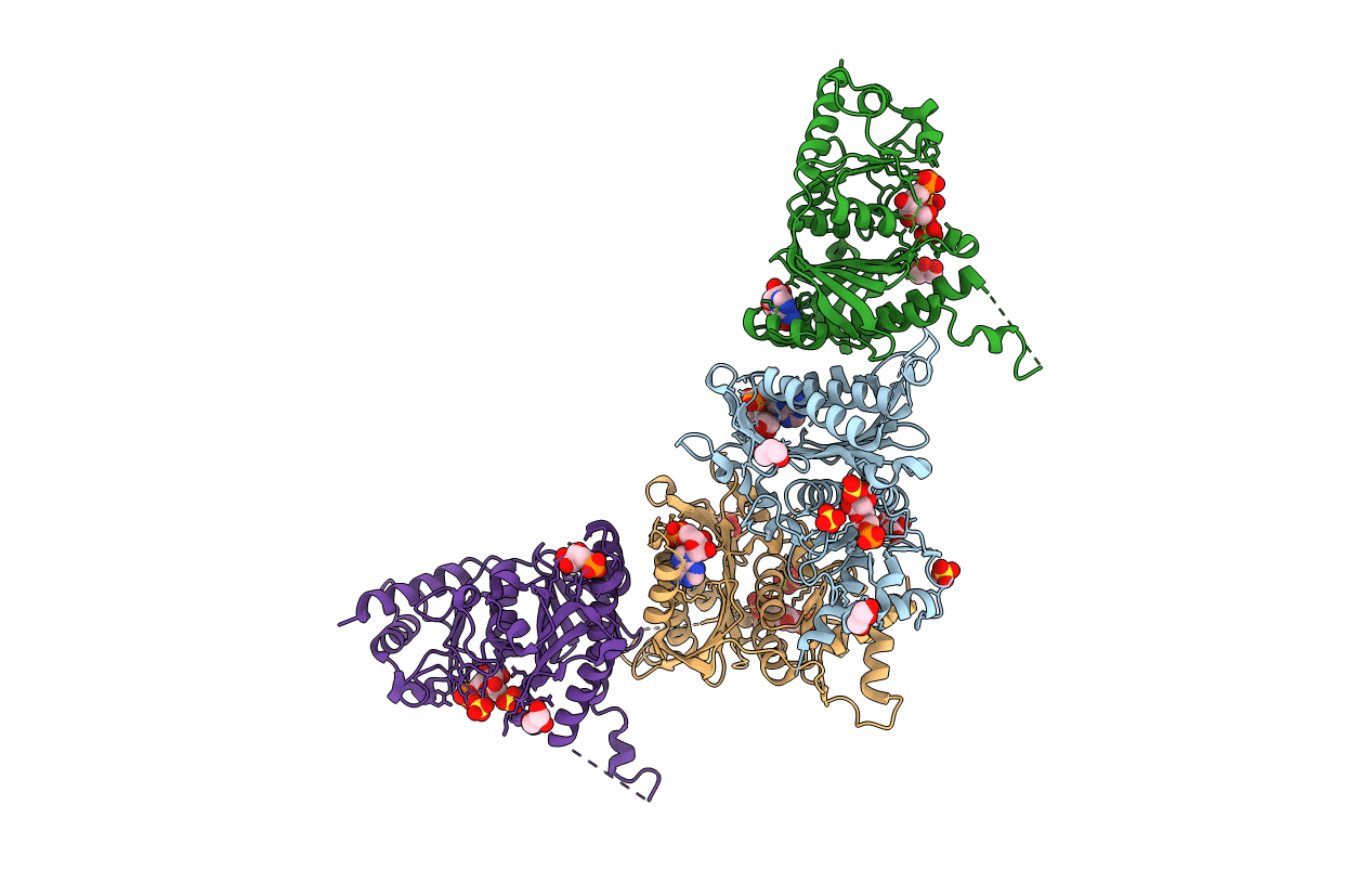
Human muscle fructose-1,6-bisphosphatase E69Q mutant in complex with AMP and alpha fructose-6-phosphate
Biological Source:
Source Organism:
Host Organism:
PDB Version:
Deposition Date:
2009-07-24
Release Date:
2010-08-11
Method Details:
Experimental Method:
Resolution:
1.97 Å
R-Value Free:
0.19
R-Value Work:
0.16
R-Value Observed:
0.16
Space Group:
C 2 2 2
Macromolecular Entities
Polymer Type:polypeptide(L)
Description:Fructose-1,6-bisphosphatase isozyme 2
Mutations:E69Q
Chain IDs:A, B, C, D
Chain Length:338
Number of Molecules:4
Biological Source:Homo sapiens
Primary Citation
Structure of E69Q mutant of human muscle fructose-1,6-bisphosphatase
Acta Crystallogr.,Sect.D 67 1028 1034 (2011)
PMID: 22120740 DOI: 10.1107/S090744491104385X

Abstact

Human fructose-1,6-bisphosphatase is an allosteric enzyme that is regulated by different ligands. There are only two known isozymes in human tissues: the liver isozyme (the key enzyme of gluconeogenesis), which is regulated by fructose 2,6-bisphosphate, and its muscle counterpart (participating in glycogen synthesis), which is regulated by calcium ions. AMP, which is an allosteric inhibitor of both isozymes, inhibits the muscle isozyme with an I(0.5) that is 35-100 times lower than for the liver isozyme and the reason for this difference remains obscure. In studies aiming at an explanation of the main differences in the regulation of the two isozymes, it has been shown that only one residue, in position 69, regulates the sensitivity towards calcium ions. As a consequence of this finding, an E69Q mutant of the muscle isozyme, which is insensitive to calcium ions while retaining all other kinetic properties resembling the liver isozyme, has been prepared and crystallized. Here, two crystal structures of this mutant enzyme in complex with AMP with and without fructose 6-phosphate (the product of the catalytic reaction) are presented. The AMP binding pattern of the muscle isozyme is quite similar to that of the liver isozyme and the T conformations of the two isozymes are nearly the same.

Legend

Protein

Chemical

Disease

Primary Citation of related structures