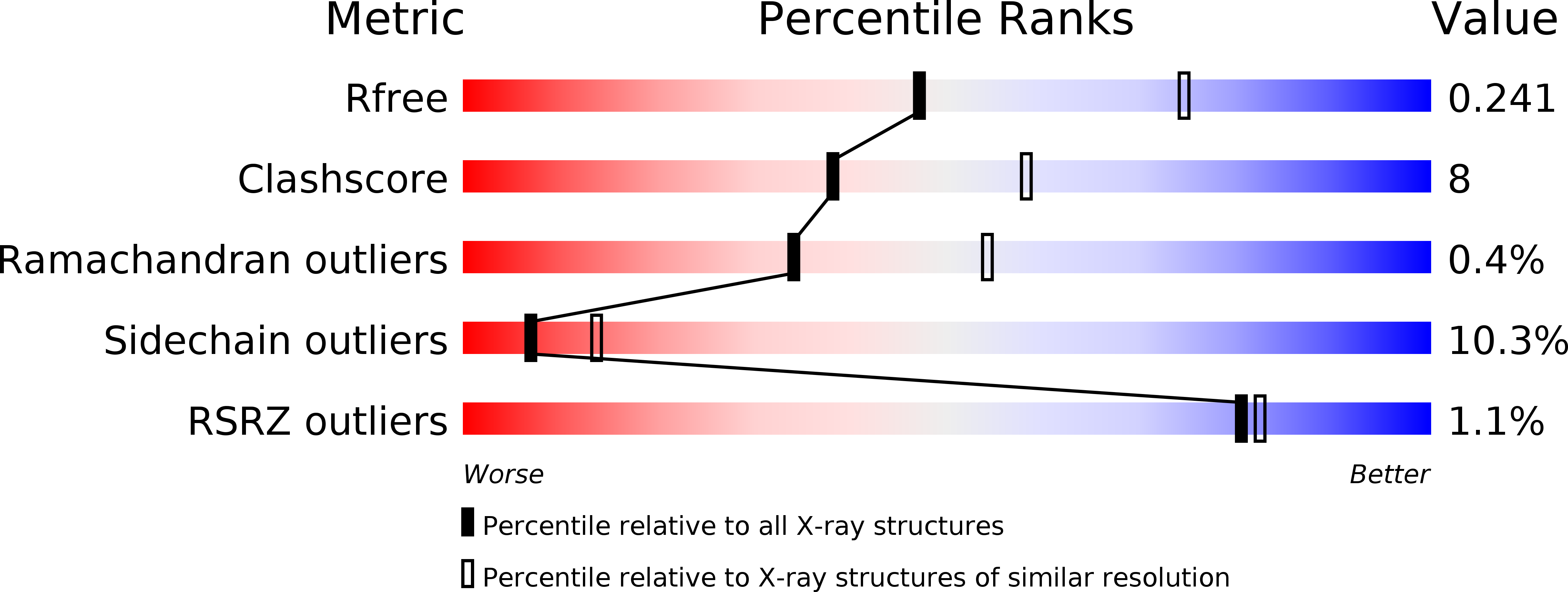
3IEG image
Deposition Date 2009-07-22
Release Date 2010-03-31
Last Version Date 2024-10-30
Entry Detail
PDB ID:
3IEG
Keywords:
Title:
Crystal Structure of P58(IPK) TPR Domain at 2.5 A
Biological Source:
Source Organism:
Mus musculus (Taxon ID: 10090)
Host Organism:
Method Details:
Experimental Method:
Resolution:
2.51 Å
R-Value Free:
0.28
R-Value Work:
0.24
R-Value Observed:
0.24
Space Group:
P 1 21 1
Macromolecular Entities
Structures with similar UniProt ID
Protein Blast
Polymer Type:polypeptide(L)
Molecule:DnaJ homolog subfamily C member 3
Gene (Uniprot):Dnajc3
Chain IDs:A, B
Chain Length:359
Number of Molecules:2
Biological Source:Mus musculus
Modified Residue
Compound ID Chain ID Parent Comp ID Details 2D Image
MSE A MET SELENOMETHIONINE
Primary Citation
Crystal Structure of P58(IPK) TPR Fragment Reveals the Mechanism for its Molecular Chaperone Activity in UPR.
J.Mol.Biol. 64 108 110 (2010)
PMID: 20184891 DOI: 10.1016/j.jmb.2010.02.028

Abstact

P58(IPK) might function as an endoplasmic reticulum molecular chaperone to maintain protein folding homeostasis during unfolded protein responses. P58(IPK) contains nine tetratricopeptide repeat (TPR) motifs and a C-terminal J-domain within its primary sequence. To investigate the mechanism by which P58(IPK) functions to promote protein folding within the endoplasmic reticulum, we have determined the crystal structure of P58(IPK) TPR fragment to 2.5 A resolution by the SAD method. The crystal structure of P58(IPK) revealed three domains (I-III) with similar folds and each domain contains three TPR motifs. An ELISA assay indicated that P58(IPK) acts as a molecular chaperone by interacting with misfolded proteins such as luciferase and rhodanese. The P58(IPK) structure reveals a conserved hydrophobic patch located in domain I that might be involved in binding the misfolded polypeptides. Structure-based mutagenesis for the conserved hydrophobic residues located in domain I significantly reduced the molecular chaperone activity of P58(IPK).

Legend

Protein

Chemical

Disease

Primary Citation of related structures
Feedback Form
Name
Email
Institute
Feedback