3IE5 image
Entry Detail
PDB ID:
3IE5
Title:
Crystal structure of Hyp-1 protein from Hypericum perforatum (St John's wort) involved in hypericin biosynthesis
Biological Source:
Source Organism:
Host Organism:
PDB Version:
Deposition Date:
2009-07-22
Release Date:
2009-11-10
Method Details:
Experimental Method:
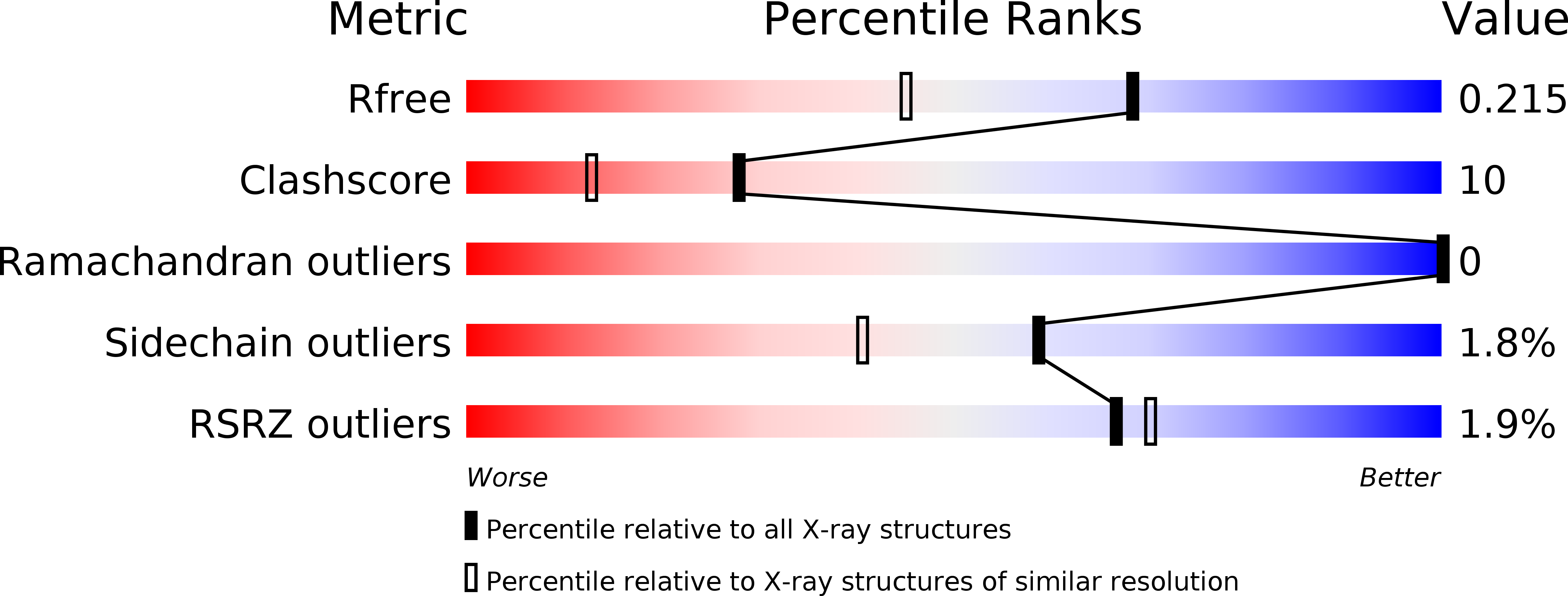
Resolution:
1.69 Å
R-Value Free:
0.20
R-Value Work:
0.17
R-Value Observed:
0.17
Space Group:
P 21 21 21
Macromolecular Entities
Polymer Type:polypeptide(L)
Description:Phenolic oxidative coupling protein Hyp-1
Chain IDs:A, B
Chain Length:165
Number of Molecules:2
Biological Source:Hypericum perforatum
Primary Citation

Abstact

Hypericin, a red-colored naphtodianthrone, is a natural product synthesized in the medicinal plant Hypericum perforatum, widely known as St. John's wort. Hypericin has been attracting a growing attention of the pharmaceutical industry because of its potential application in various therapies, including the treatment of depression. In vivo, hypericin is synthesized by dimerization of emodin in a complicated multistep reaction that is reportedly catalyzed by a small (17.8kDa) protein, Hyp-1. Based on relatively low sequence similarity (approximately 50%), Hyp-1 has been tentatively classified as a plant PR-10 (pathogenesis-related class 10) protein. Members of the PR-10 family are ubiquitous plant proteins associated with stress control and tissue differentiation but with no clearly understood molecular mechanism. They have, however, a well-defined folding canon, consisting of an extended antiparallel beta-sheet wrapped around a C-terminal alpha-helix, enclosing in the protein interior a huge cavity, in which various hydrophobic ligands can be bound. Apart from Hyp-1, only two other PR-10 members have been found to possess enzymatic activity (S-norcoclaurine synthase and TcmN aromatase/cyclase). In this paper, we report a high-resolution crystal structure of Hyp-1, confirming that it indeed has a PR-10 fold. The protein binds multiple polyethylene glycol molecules, some of which occupy the hydrophobic cavity. The crystallographic model illustrates a high degree of conformational adaptability of both interacting partners for efficient binding. We have been unable, however, to dimerize emodin to hypericin using Hyp-1 as biocatalyst. This puzzling result does not have a clear explanation at this time.

Legend

Protein

Chemical

Disease

Primary Citation of related structures