3ID7 image
Entry Detail
PDB ID:
3ID7
Keywords:
Title:
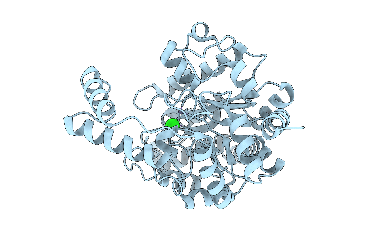
Crystal structure of renal dipeptidase from Streptomyces coelicolor A3(2)
Biological Source:
Source Organism:
Host Organism:
PDB Version:
Deposition Date:
2009-07-20
Release Date:
2010-01-12
Method Details:
Experimental Method:
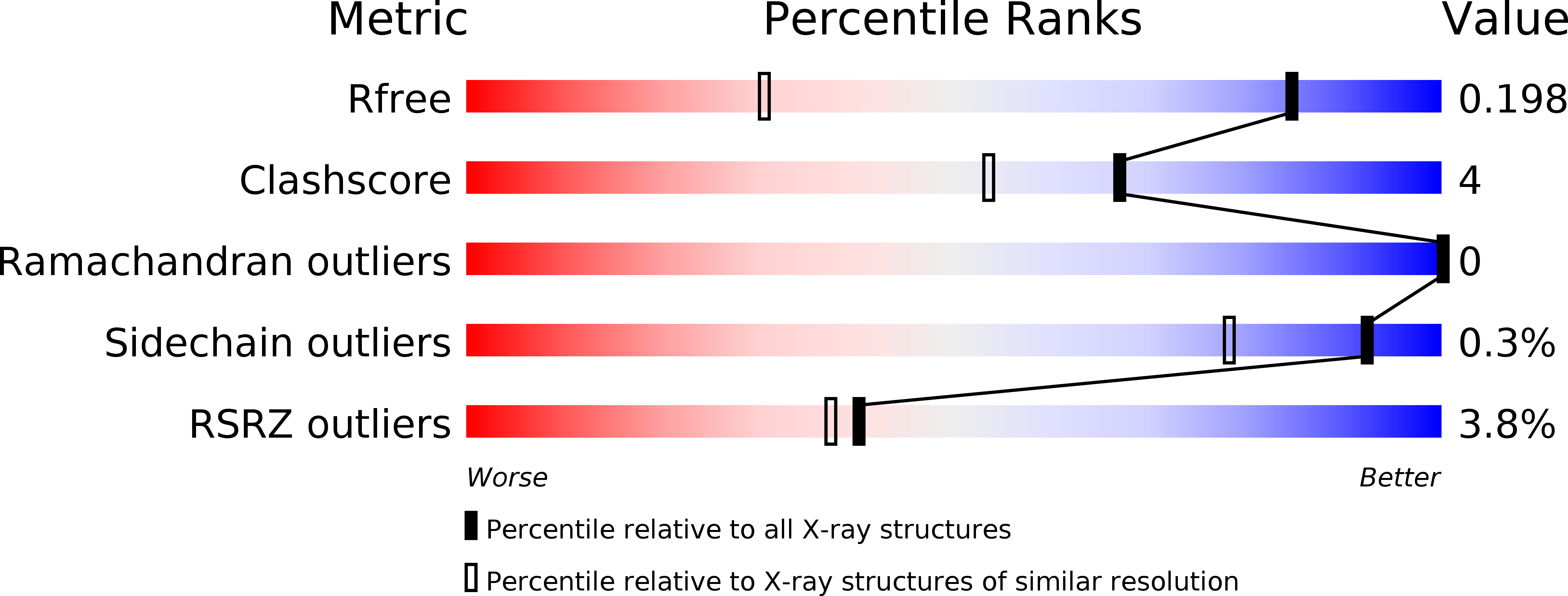
Resolution:
1.30 Å
R-Value Free:
0.19
R-Value Work:
0.18
R-Value Observed:
0.18
Space Group:
P 31 2 1
Macromolecular Entities
Polymer Type:polypeptide(L)
Description:Dipeptidase
Chain IDs:A
Chain Length:400
Number of Molecules:1
Biological Source:Streptomyces coelicolor
Primary Citation
Structure, mechanism, and substrate profile for Sco3058: the closest bacterial homologue to human renal dipeptidase .
Biochemistry 49 611 622 (2010)
PMID: 20000809 DOI: 10.1021/bi901935y

Abstact

Human renal dipeptidase, an enzyme associated with glutathione metabolism and the hydrolysis of beta-lactams, is similar in sequence to a cluster of approximately 400 microbial proteins currently annotated as nonspecific dipeptidases within the amidohydrolase superfamily. The closest homologue to the human renal dipeptidase from a fully sequenced microbe is Sco3058 from Streptomyces coelicolor. Dipeptide substrates of Sco3058 were identified by screening a comprehensive series of l-Xaa-l-Xaa, l-Xaa-d-Xaa, and d-Xaa-l-Xaa dipeptide libraries. The substrate specificity profile shows that Sco3058 hydrolyzes a broad range of dipeptides with a marked preference for an l-amino acid at the N-terminus and a d-amino acid at the C-terminus. The best substrate identified was l-Arg-d-Asp (k(cat)/K(m) = 7.6 x 10(5) M(-1) s(-1)). The three-dimensional structure of Sco3058 was determined in the absence and presence of the inhibitors citrate and a phosphinate mimic of l-Ala-d-Asp. The enzyme folds as a (beta/alpha)(8) barrel, and two zinc ions are bound in the active site. Site-directed mutagenesis was used to probe the importance of specific residues that have direct interactions with the substrate analogues in the active site (Asp-22, His-150, Arg-223, and Asp-320). The solvent viscosity and kinetic effects of D(2)O indicate that substrate binding is relatively sticky and that proton transfers do not occurr during the rate-limiting step. A bell-shaped pH-rate profile for k(cat) and k(cat)/K(m) indicated that one group needs to be deprotonated and a second group must be protonated for optimal turnover. Computational docking of high-energy intermediate forms of l/d-Ala-l/d-Ala to the three-dimensional structure of Sco3058 identified the structural determinants for the stereochemical preferences for substrate binding and turnover.

Legend

Protein

Chemical

Disease

Primary Citation of related structures