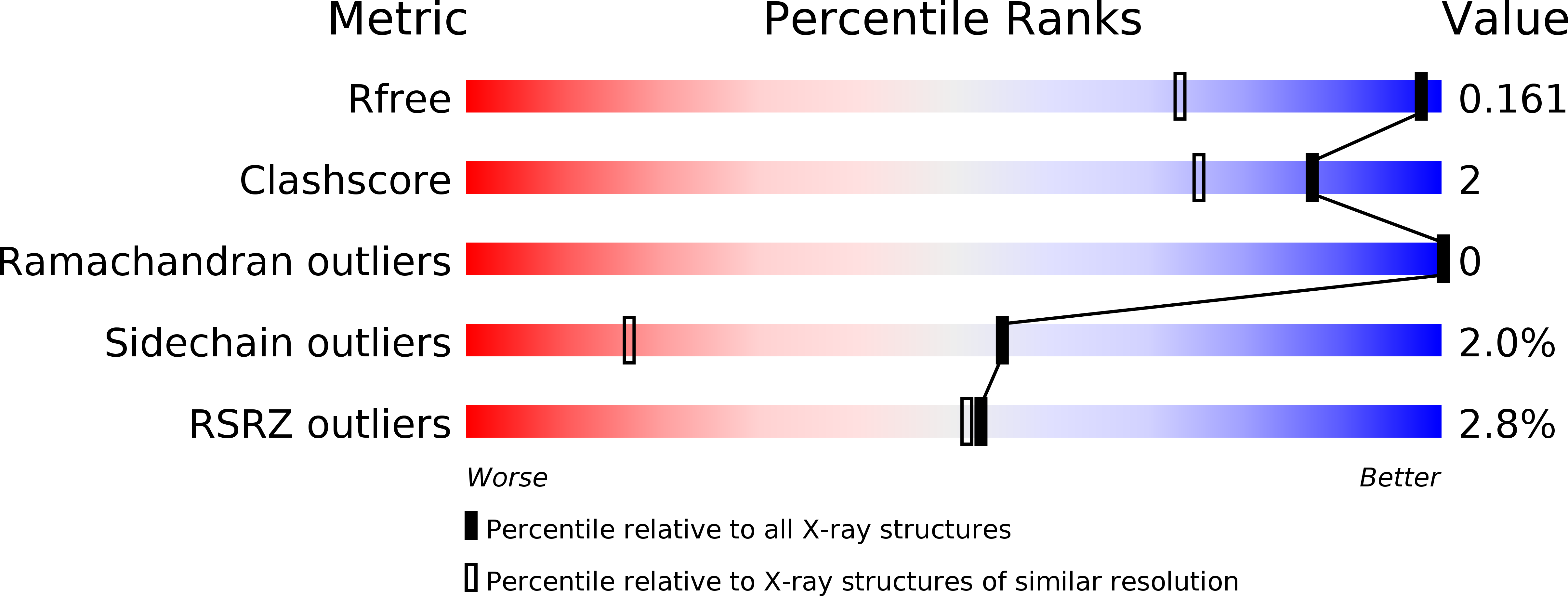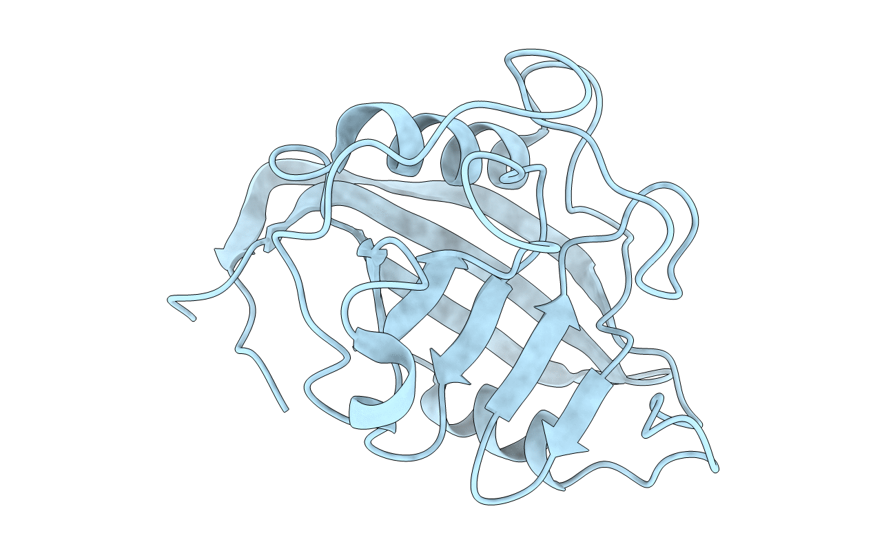
3ICH image
Deposition Date 2009-07-17
Release Date 2010-07-21
Last Version Date 2023-09-06
Entry Detail
PDB ID:
3ICH
Keywords:
Title:
Crystal structure of cyclophilin B at 1.2 A resolution
Biological Source:
Source Organism:
Homo sapiens (Taxon ID: 9606)
Host Organism:
Method Details:
Experimental Method:
Resolution:
1.20 Å
R-Value Free:
0.16
R-Value Work:
0.14
R-Value Observed:
0.14
Space Group:
P 32
Macromolecular Entities
Polymer Type:polypeptide(L)
Molecule:Peptidyl-prolyl cis-trans isomerase B
Gene (Uniprot):PPIB
Chain IDs:A
Chain Length:188
Number of Molecules:1
Biological Source:Homo sapiens
Primary Citation
Structural Basis of Cyclophilin B Binding by the Calnexin/Calreticulin P-domain.
J.Biol.Chem. 285 35551 35557 (2010)
PMID: 20801878 DOI: 10.1074/jbc.M110.160101

Abstact

Little is known about how chaperones in the endoplasmic reticulum are organized into complexes to assist in the proper folding of secreted proteins. One notable exception is the complex of ERp57 and calnexin that functions as part the calnexin cycle to direct disulfide bond formation in N-glycoproteins. Here, we report three new complexes composed of the peptidyl prolyl cis-trans-isomerase cyclophilin B and any of the lectin chaperones: calnexin, calreticulin, or calmegin. The 1.7 Å crystal structure of cyclophilin with the proline-rich P-domain of calmegin reveals that binding is mediated by the same surface that binds ERp57. We used NMR titrations and mutagenesis to measure low micromolar binding of cyclophilin to all three lectin chaperones and identify essential interfacial residues. The immunosuppressant cyclosporin A did not affect complex formation, confirming the functional independence of the P-domain binding and proline isomerization sites of cyclophilin. Our results reveal the P-domain functions as a unique protein-protein interaction domain and implicate a peptidyl prolyl isomerase as a new element in the calnexin cycle.

Legend

Protein

Chemical

Disease

Primary Citation of related structures