3IBO image
Entry Detail
PDB ID:
3IBO
Title:
Pseudomonas aeruginosa E2Q/H83Q/T126H-azurin RE(PHEN)(CO)3
Biological Source:
Source Organism:
Host Organism:
PDB Version:
Deposition Date:
2009-07-16
Release Date:
2009-12-08
Method Details:
Experimental Method:
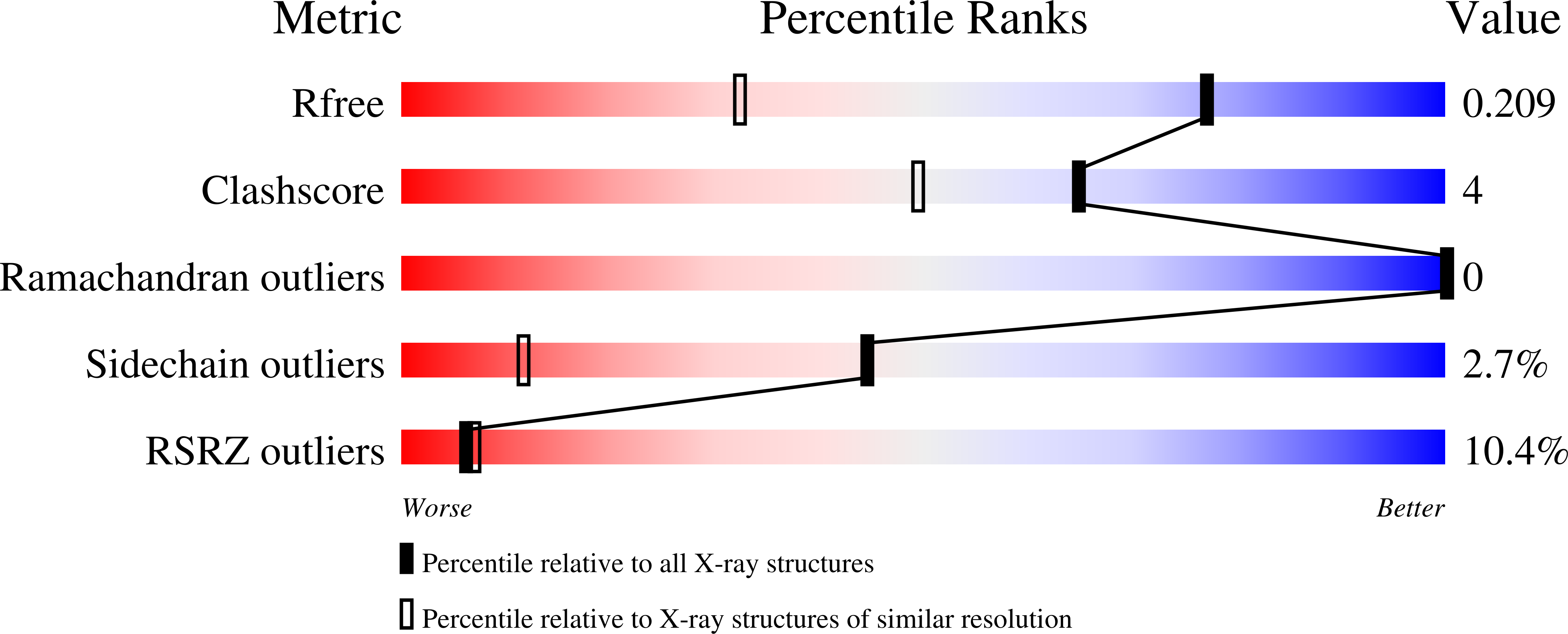
Resolution:
1.45 Å
R-Value Free:
0.21
R-Value Work:
0.20
Space Group:
P 1 21 1
Macromolecular Entities
Polymer Type:polypeptide(L)
Description:Azurin
Mutations:E2Q, H83Q, T126H
Chain IDs:A, B, C, D
Chain Length:128
Number of Molecules:4
Biological Source:Pseudomonas aeruginosa
Primary Citation
Relaxation dynamics of Pseudomonas aeruginosa Re(I)(CO)3(alpha-diimine)(HisX)+ (X = 83, 107, 109, 124, 126)Cu(II) azurins.
J.Am.Chem.Soc. 131 11788 11800 (2009)
PMID: 19639996 DOI: 10.1021/ja902744s

Abstact

Photoinduced relaxation processes of five structurally characterized Pseudomonas aeruginosa Re(I)(CO)(3)(alpha-diimine)(HisX) (X = 83, 107, 109, 124, 126)Cu(II) azurins have been investigated by time-resolved (ps-ns) IR spectroscopy and emission spectroscopy. Crystal structures reveal the presence of Re-azurin dimers and trimers that in two cases (X = 107, 124) involve van der Waals interactions between interdigitated diimine aromatic rings. Time-dependent emission anisotropy measurements confirm that the proteins aggregate in mM solutions (D(2)O, KP(i) buffer, pD = 7.1). Excited-state DFT calculations show that extensive charge redistribution in the Re(I)(CO)(3) --> diimine (3)MLCT state occurs: excitation of this (3)MLCT state triggers several relaxation processes in Re-azurins whose kinetics strongly depend on the location of the metallolabel on the protein surface. Relaxation is manifested by dynamic blue shifts of excited-state nu(CO) IR bands that occur with triexponential kinetics: intramolecular vibrational redistribution together with vibrational and solvent relaxation give rise to subps, approximately 2, and 8-20 ps components, while the approximately 10(2) ps kinetics are attributed to displacement (reorientation) of the Re(I)(CO)(3)(phen)(im) unit relative to the peptide chain, which optimizes Coulombic interactions of the Re(I) excited-state electron density with solvated peptide groups. Evidence also suggests that additional segmental movements of Re-bearing beta-strands occur without perturbing the reaction field or interactions with the peptide. Our work demonstrates that time-resolved IR spectroscopy and emission anisotropy of Re(I) carbonyl-diimine complexes are powerful probes of molecular dynamics at or around the surfaces of proteins and protein-protein interfacial regions.

Legend

Protein

Chemical

Disease

Primary Citation of related structures