3I6K image
Deposition Date 2009-07-07
Release Date 2010-06-16
Last Version Date 2024-10-30
Entry Detail
PDB ID:
3I6K
Keywords:
Title:
Newly identified epitope from SARS-CoV membrane protein complexed with HLA-A*0201
Biological Source:
Source Organism:
Host Organism:
Method Details:
Experimental Method:
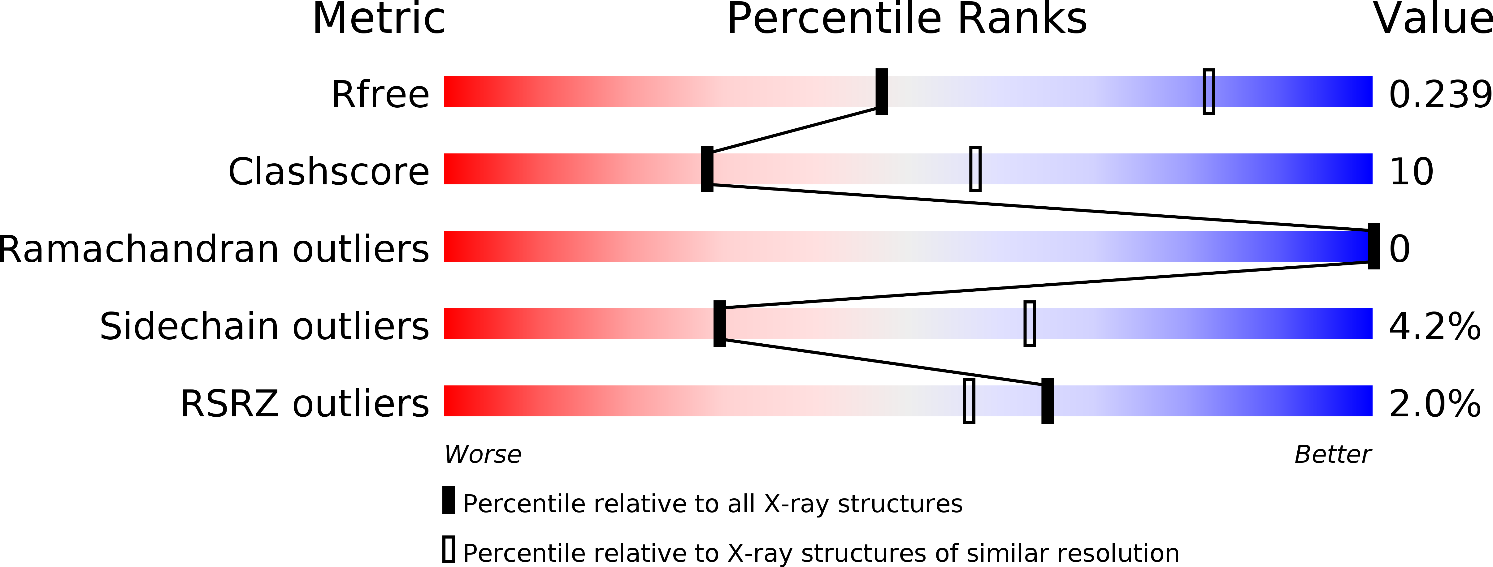
Resolution:
2.80 Å
R-Value Free:
0.24
R-Value Work:
0.19
R-Value Observed:
0.19
Space Group:
P 21 21 21
Macromolecular Entities
Structures with similar UniProt ID
Protein Blast
Polymer Type:polypeptide(L)
Molecule:HLA class I histocompatibility antigen, A-2 alpha chain
Chain IDs:A, D (auth: E)
Chain Length:275
Number of Molecules:2
Biological Source:Homo sapiens
Structures with similar UniProt ID
Protein Blast
Polymer Type:polypeptide(L)
Molecule:Beta-2-microglobulin
Gene (Uniprot):B2M
Chain IDs:B, E (auth: F)
Chain Length:100
Number of Molecules:2
Biological Source:Homo sapiens
Structures with similar UniProt ID
Protein Blast
Polymer Type:polypeptide(L)
Molecule:Membrane glycoprotein peptide
Chain IDs:C, F (auth: G)
Chain Length:10
Number of Molecules:2
Biological Source:SARS coronavirus TJF
Primary Citation
The membrane protein of severe acute respiratory syndrome coronavirus acts as a dominant immunogen revealed by a clustering region of novel functionally and structurally defined cytotoxic T-lymphocyte epitopes
J Infect Dis 202 1171 1180 (2010)
PMID: 20831383 DOI: 10.1086/656315

Abstact

BACKGROUND Severe acute respiratory syndrome coronavirus (SARS-CoV), which emerged with highly contagious and life-threatening characteristics in 2002, remains a potential risk for future outbreaks. Membrane (M) and envelope (E) proteins are major structural proteins of the SARS-CoV. The M protein has been determined as a protective antigen in humoral responses. However, its potential roles in stimulating cellular immunity remain elusive. METHODS In this study, a panel of peptides derived from M and E proteins were tested by in vitro refolding, T2 cell-binding assays, and responses stimulated by cytotoxic T-lymphocyte (CTL) epitopes in HLA-A2.1/K(b) transgenic mice and human peripheral blood mononuclear cells (PBMCs). RESULTS A nonameric epitope Mn2 and a decameric epitope Md3 derived from the M protein were identified and used for the evaluation of M protein-specific immunity. Responses stimulated by M protein-specific CTL epitopes have been found in the PBMCs of donors who had recovered from SARS infection. Additionally, the transmembrane domain of the M protein may contain a T cell epitope cluster revealed by the immunogenic and structural analysis of a panel of truncated peptides overlapping with Mn2 and Md3. CONCLUSIONS The M protein of SARS-CoV holds dominant cellular immunogenicity. This, together with previous reports of a strong humoral response against the M protein, may help to further explain the immunogenicity of SARS and serves as potential targets for SARS-CoV vaccine design.

Legend

Protein

Chemical

Disease

Primary Citation of related structures