Deposition Date
2009-06-25
Release Date
2009-07-28
Last Version Date
2023-09-06
Entry Detail
PDB ID:
3I17
Keywords:
Title:
Crystal structure of the apo R132K:L121E mutant of cellular retinoic acid-binding protein II at 1.68 angstrom resolution
Biological Source:
Source Organism(s):
Homo sapiens (Taxon ID: 9606)
Expression System(s):
Method Details:
Experimental Method:
Resolution:
1.68 Å
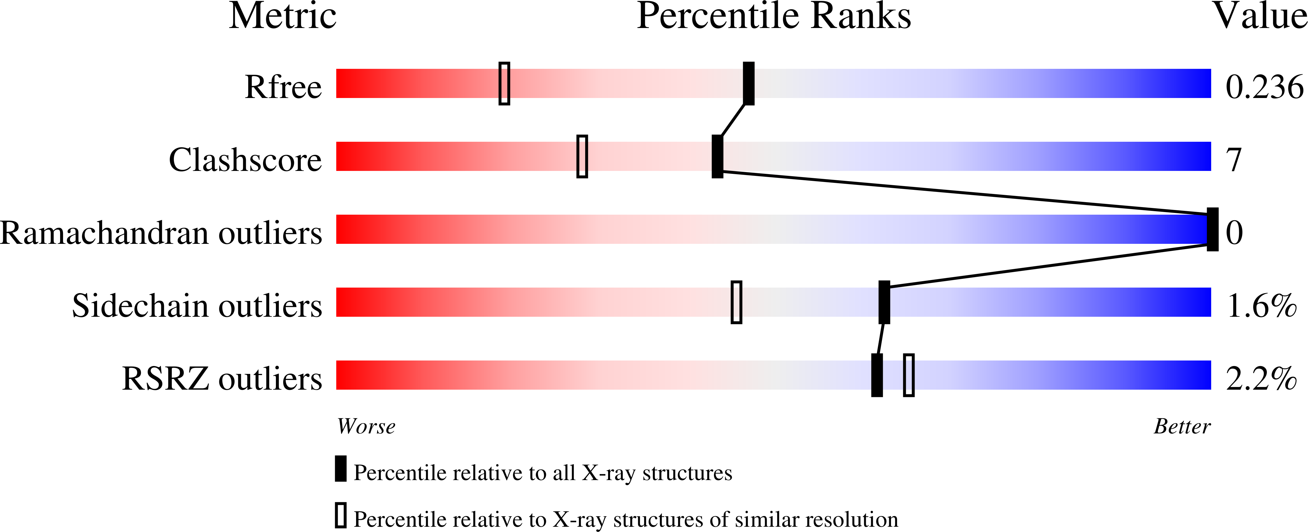
R-Value Free:
0.23
R-Value Work:
0.18
R-Value Observed:
0.18
Space Group:
P 1