Deposition Date
2009-06-23
Release Date
2010-02-16
Last Version Date
2024-11-27
Entry Detail
PDB ID:
3HZH
Keywords:
Title:
Crystal structure of the CheX-CheY-BeF3-Mg+2 complex from Borrelia burgdorferi
Biological Source:
Source Organism(s):
Borrelia burgdorferi (Taxon ID: 139)
Expression System(s):
Method Details:
Experimental Method:
Resolution:
1.96 Å
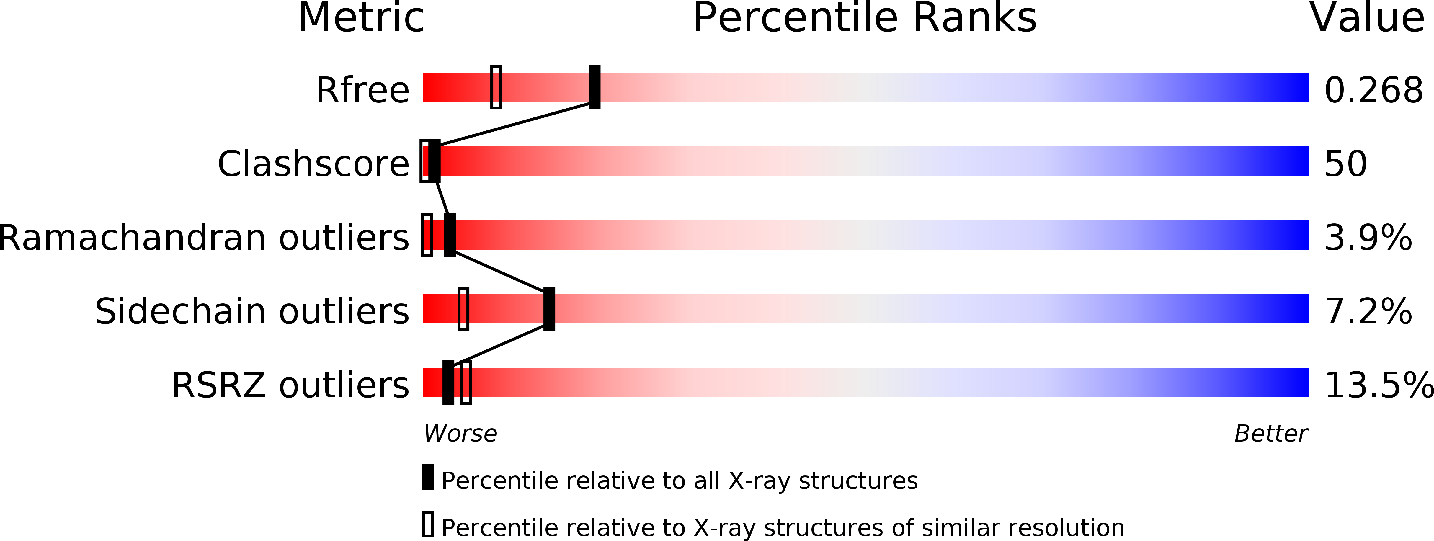
R-Value Free:
0.25
R-Value Work:
0.24
Space Group:
P 32 2 1