
Deposition Date
2009-06-19
Release Date
2009-12-01
Last Version Date
2024-02-21
Entry Detail
PDB ID:
3HX9
Keywords:
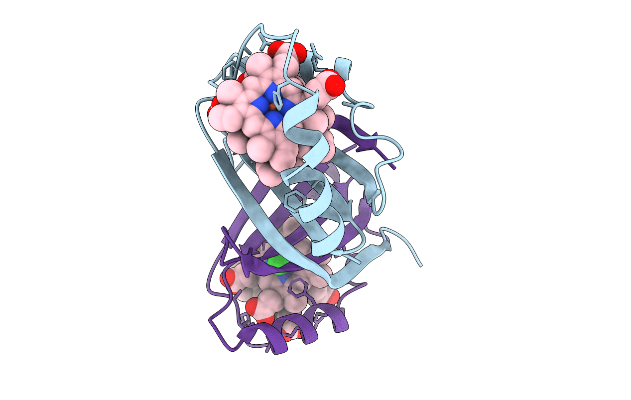
Title:
Structure of heme-degrader, MhuD (Rv3592), from Mycobacterium tuberculosis with two hemes bound in its active site
Biological Source:
Source Organism(s):
Mycobacterium tuberculosis (Taxon ID: 1773)
Expression System(s):
Method Details:
Experimental Method:
Resolution:
1.75 Å
R-Value Free:
0.23
R-Value Work:
0.18
R-Value Observed:
0.18
Space Group:
C 1 2 1
